Rice stripe virus immune detection reagent kit

A technology for rice streak virus and immune detection, applied in the field of agricultural science, can solve the problems of high cost and inappropriate detection of samples in large quantities

Inactive Publication Date: 2006-09-06
JIANGSU ACAD OF AGRI SCI
View PDF0 Cites 9 Cited by
  • Summary
  • Abstract
  • Description
  • Claims
  • Application Information

AI Technical Summary

Problems solved by technology

At present, the methods used in the world to detect the infection rate of SBPH include ELISA, RT-PCR, Northern hybridization, etc., but RT-PCR and Northern hybridization are expensive and not suitable for large-scale detection of samples; Zhou Yijun et al. prepared RSV monoclonal antibody for virus Detection, based on this, the RSV detection method established by dot immunoassay is fast, sensitive, simple and easy to implement, and it is expected to detect a kind of RSV detection technology suitable for the application of grassroots agricultural technicians in my country, and carry out early detection of poisonous insects in the early stage And timely warning, and can also be applied to the basic field of research on the interaction between RSV, SBPH and plant hosts

Method used


Image

  • Rice stripe virus immune detection reagent kit
  • Rice stripe virus immune detection reagent kit
  • Rice stripe virus immune detection reagent kit

Examples


Embodiment 1

[0046] To detect the virus-carrying condition of the transmission medium SBPH, the steps at room temperature are as follows:

[0047] 1) Dilute the washing solution 10 times to 0.01M with pure water, and prepare the blocking solution for later use.

[0048] 2) Draw 0.25cm on the nitrocellulose membrane (NC) membrane with a pencil 2 left and right squares.

[0049] 3) Add 2 drops of sample treatment solution to a 0.2ml centrifuge tube, mash it with a toothpick, take a small drop of supernatant (about 3 μL) and apply it to the center of the nitrocellulose membrane grid, and dry it at room temperature.

[0050] 4) Immerse the dried membrane in blocking solution for 1 h at room temperature.

[0051] 5) Add one unit of monoclonal antibody to 10 ml of blocking solution, take out the membrane and immerse it, and place it at room temperature for 3 hours.

[0052] 6) Take out the membrane and wash with lotion 3 times, each time for 3-5 minutes.

[0053] 7) Add one unit of secondary...

Embodiment 2

[0059] Sample dilution test

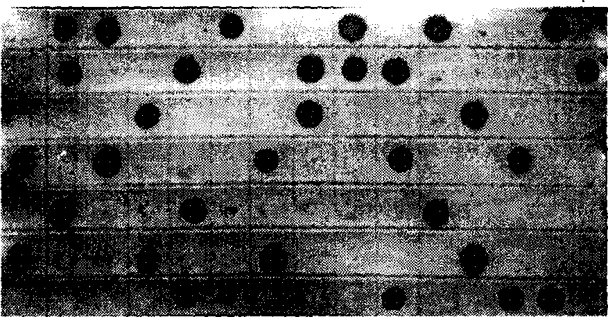
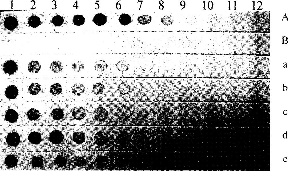
[0060] Single-headed SBPH and diseased leaves were diluted into 11 gradients starting from 100 times, and the non-toxic SBPH was used as a negative control at the same dilution.

[0061]The results show that a weak positive reaction can still be seen when diluted to 25,600 times, but the strongest positive reaction can be seen at a dilution of 100 times, and a dilution of 100-200 times is usually used for routine detection; weak positive reactions can still be seen when the diseased leaves are diluted to 51,200 times , but 400-fold dilution is better for visual inspection, and 200-500-fold dilution ( figure 2 ). But it is best to use fresh diseased leaves or use the method of detecting plant tissue.

Embodiment 3

[0063] Antibody Incubation Time Test

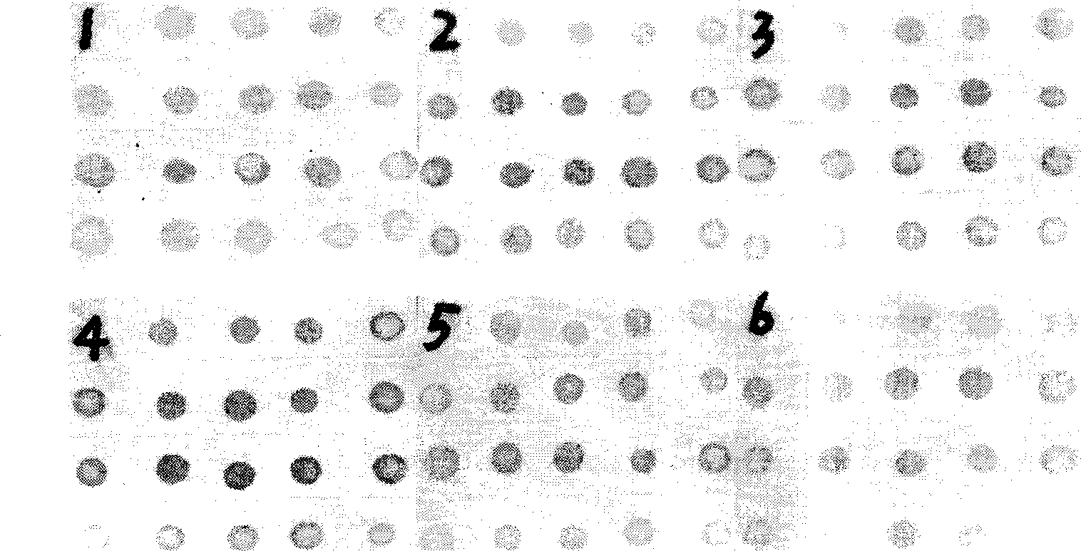
[0064] Monoclonal antibody working concentration 1:20000, incubate at 37°C for 0.5h, 1h, 1.5h, 2h, 2.5h, 3h (secondary antibody concentration 1:2500, secondary antibody incubation 1.5h) to determine the change of monoclonal antibody incubation time impact on test results. The results showed that when incubated at 37°C for 0.5h, positive samples could be detected, the color of the spots when incubated for 1h was basically the same as when incubated for 1.5h, 2h, and 2.5h, and the color decreased slightly when incubated for 3h ( image 3 ). Therefore, it is determined that incubation of monoclonal antibody at 37°C for 1-1.5h is the best incubation time.

[0065] Determine the monoclonal antibody concentration of 1:20000, and incubate at 37°C for 1.5h; use the concentration of secondary antibody at 1:2500, and incubate at 37°C for 0.5h, 1h, 1.5h, 2h, 2.5h, and 3h, respectively, to determine whether to change the secondary antibody Effect ...


PUM


Abstract

Wherein, building a DIPA detection method with the RSV specific monoclonal antibody, and hereby building the reagent box with main reagent provided as working liquid or condensed liquid. This invention can detect whether there is RSV rapidly, and has wide application.

Description

[0001] The invention relates to an immune detection kit for rice stripe virus and its application in agriculture, belonging to the field of agricultural science and technology. Background technique [0002] Rice stripe blight caused by rice stripe virus (RSV) was first discovered in Kanto, Japan in 1897, and later occurred in Korea, Ukraine and China. Since the virus was discovered in Jiangsu and Zhejiang in 1962 in my country, it has been prevalent in 16 provinces, municipalities and autonomous regions across the country. The disease is still very common in Yunnan, Liaoning, Beijing, Henan, Shandong, Jiangsu, and Shanghai, especially Baoshan, Chuxiong, and Kunming in Yunnan, Shuangqiao in Beijing, Yuanyang in Henan, Jining in Shandong, and Jiangyan and Hong in northern Jiangsu. Ze and other places, the occurrence is more common. [0003] The main reasons for its continuous outbreak are: the warm winter climate in recent years is conducive to the overwintering of the virus-tr...

Claims


Application Information

Patent Timeline
Patent Type & Authority Applications(China)
IPC IPC(8): G01N33/577G01N33/569
Inventor 周益军周彤程兆榜熊如意刘海建
Owner JIANGSU ACAD OF AGRI SCI
Why Eureka
Try Eureka