X-ray computed tomography apparatus

A tomography and X-ray technology, applied in computer tomography scanners, instruments for radiological diagnosis, diagnosis, etc., can solve problems such as reconstruction, confirmation, and deterioration of reconstructed image quality.

Active Publication Date: 2007-03-14
TOSHIBA MEDICAL SYST CORP
View PDF4 Cites 18 Cited by
  • Summary
  • Abstract
  • Description
  • Claims
  • Application Information

AI Technical Summary

Problems solved by technology

[0009] However, due to arrhythmia, etc., sometimes the projection data required for image reconstruction of the desired heartbeat time phase is not collected, or the projection data is not collected at a high SN ratio.
In this case, the quality of the reconstructed image deteriorates
Depending on the situation, sometimes a rescan is necessary
Also, there is no technique to confirm the above before rebuilding

Method used


Image

  • X-ray computed tomography apparatus
  • X-ray computed tomography apparatus
  • X-ray computed tomography apparatus

Examples


Embodiment approach 1

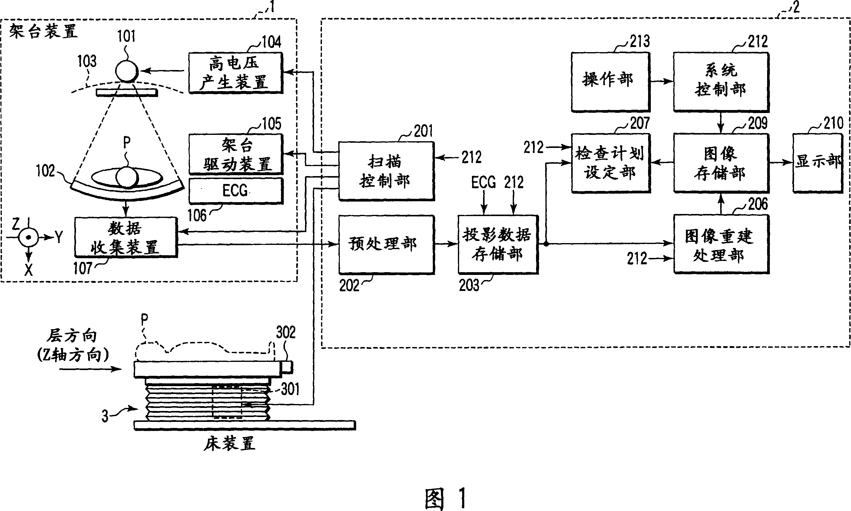
[0039] Next, Embodiment 1 of the X-ray computed tomography apparatus of the present invention will be described with reference to the drawings. X-ray computed tomography apparatus includes: a rotation / rotation (ROTATE / ROTATE) type in which the X-ray tube and the X-ray detector are integrally rotated around the subject; The present invention can be applied to any of various types such as the STATIONARY / ROTATE type in which the X-ray tube rotates around the object to be examined. Here, it will be described as a rotary / rotary type currently in the mainstream. In addition, in order to reconstruct the tomographic image data of one slice (slice), projection data of about 360 degrees around the subject are required (full reconstruction method), and even the half reconstruction method requires 180 degrees + α (α : projection data of the size of fan angle). In this embodiment, a semi-reconstruction method effective for imaging a fast-moving heart or the like is employed.

[0040] In...

Embodiment approach 2

[0138] Next, a second embodiment of the X-ray computed tomography apparatus (also referred to as X-ray CT or CT scanner) of the present invention will be described with reference to the drawings. The X-ray computed tomography apparatus includes: a rotation / rotation (ROTATE / ROTATE) type in which an X-ray tube and a radiation detector are integrally rotated around an object; The present invention can be applied to any of various types such as a stationary / rotate type that rotates around the subject. Here, it will be described as a rotary / rotary type currently in the mainstream.

[0139] In addition, reconstruction methods include full reconstruction methods, semi-reconstruction methods, and segmental reconstruction methods. These reconstruction methods differ in the range of angles required to reconstruct one-slice tomographic data. The time required to rotate the X-ray tube to reconstruct the angular range required for one slice of tomographic data is called time resolution. ...


PUM


Abstract

An X-ray computed tomography apparatus includes an X-ray source (101) which generates X-rays, a high voltage generating unit (104) which generates a high voltage to be applied to the X-ray source, an X-ray detector (102) which detects X-rays transmitted through a subject to be examined to generate projection data, a storage unit (203) which stores the projection data in association with electrocardiographic data of the subject, a setting unit (207) which sets a specific cardiac phase in accordance with an operator's instruction, a reconstruction unit (206) which reconstructs a tomogram on the basis of a plurality of projection data sets acquired in a plurality of specific periods centered on the specific cardiac phase throughout a plurality of cardiac cycles, a period extending unit (207) which extends the specific period on the basis of a heart rate fluctuation range of the subject, and a control unit (212) which controls the high voltage generating unit to generate a relatively high dose of X-rays in the extended specific period and generate a relatively low dose of X-rays in a period other than the extended specific period.

Description

[0001] Cross References to Related Applications [0002] This application is based upon and claims the benefit of priority from prior Japanese Patent Applications No. 2005-259614 filed on September 7, 2005 and No. 2005-285863 filed on September 30, 2005, the entire contents of which are incorporated herein by reference middle. technical field [0003] The present invention relates to an X-ray computed tomography device capable of synchronous reconstruction of electrocardiogram (ECG gated reconstruction: ECGgated reconstruction). Background technique [0004] X-ray computed tomography device uses images to provide information about the subject according to the intensity of the X-rays that pass through the subject, and plays an important role in various medical activities represented by disease diagnosis, treatment, and surgical planning. effect. [0005] In fast-moving cardiac exams, an electrocardiographically synchronized scan is performed. The so-called ECG synchronous ...

Claims


Application Information

Patent Timeline
Patent Type & Authority Applications(China)
IPC IPC(8): A61B6/03G01N23/04
CPCA61B6/541
Inventor 长田清津雪昌快
Owner TOSHIBA MEDICAL SYST CORP
Why Eureka
Try Eureka