Image processing system and method for detecting an anatomical marker within an image study

an image processing system and image technology, applied in image enhancement, instruments, healthcare informatics, etc., can solve the problems of requiring time-consuming and taxing review, and requiring substantial computing resources, so as to achieve the processing resources required to detect time and efficiently

Active Publication Date: 2018-09-18
CHANGE HEALTHCARE HLDG LLC
View PDF4 Cites 0 Cited by
  • Summary
  • Abstract
  • Description
  • Claims
  • Application Information

AI Technical Summary

Benefits of technology

[0006]A method, image processing system and computer program product are provided in accordance with an example embodiment in order to detect an anatomical marker within an image study. In this regard, the method, image processing system and computer program product of an example embodiment may be configured to detect an anatomical marker based upon data elements that are representative of one or more characteristics of the image slices, but that do not include picture elements. Thus, the method, image processing system and computer program product of this example embodiment may detect an anatomical marker in a more timely and efficient manner. Consequently, the processing resources required to detect an anatomical marker may be conserved, while still permitting a subset of the image slices of the image study to be reliably identified based upon the anatomical marker or a distance from the anatomical marker, such as for storage, display or the like.

Problems solved by technology

This review may be time consuming and taxing as the physician or other health care professional must attentively review numerous image slices to identify those of most interest.
As such, fiducial or anatomic landmark detection techniques are relatively complex and employ substantial computing resources in order to detect the desired fiducial or anatomic markers.
In this regard, the complexity of the fiducial or anatomic landmark detection techniques may increase depending upon the nature of the fiducial or anatomic markers with many image slices being required to be subjected to the image processing and analysis techniques.
Thus, these techniques not only require access to the values of the picture elements of the different image slices, but also generally require the processing of substantial amounts of data, such as the analysis of large numbers of pixel intensity values for model matching or other purposes.
Thus, these fiducial or anatomic landmark detection techniques require substantial processing power and utilize relatively large amounts of memory.

Method used


Image

  • Image processing system and method for detecting an anatomical marker within an image study
  • Image processing system and method for detecting an anatomical marker within an image study
  • Image processing system and method for detecting an anatomical marker within an image study

Examples


Embodiment Construction

[0019]Some embodiments of the present invention will now be described more fully hereinafter with reference to the accompanying drawings, in which some, but not all, embodiments of the invention are shown. Indeed, various embodiments of the invention may be embodied in many different forms and should not be construed as limited to the embodiments set forth herein; rather, these embodiments are provided so that this disclosure will satisfy applicable legal requirements. Like reference numerals refer to like elements throughout.

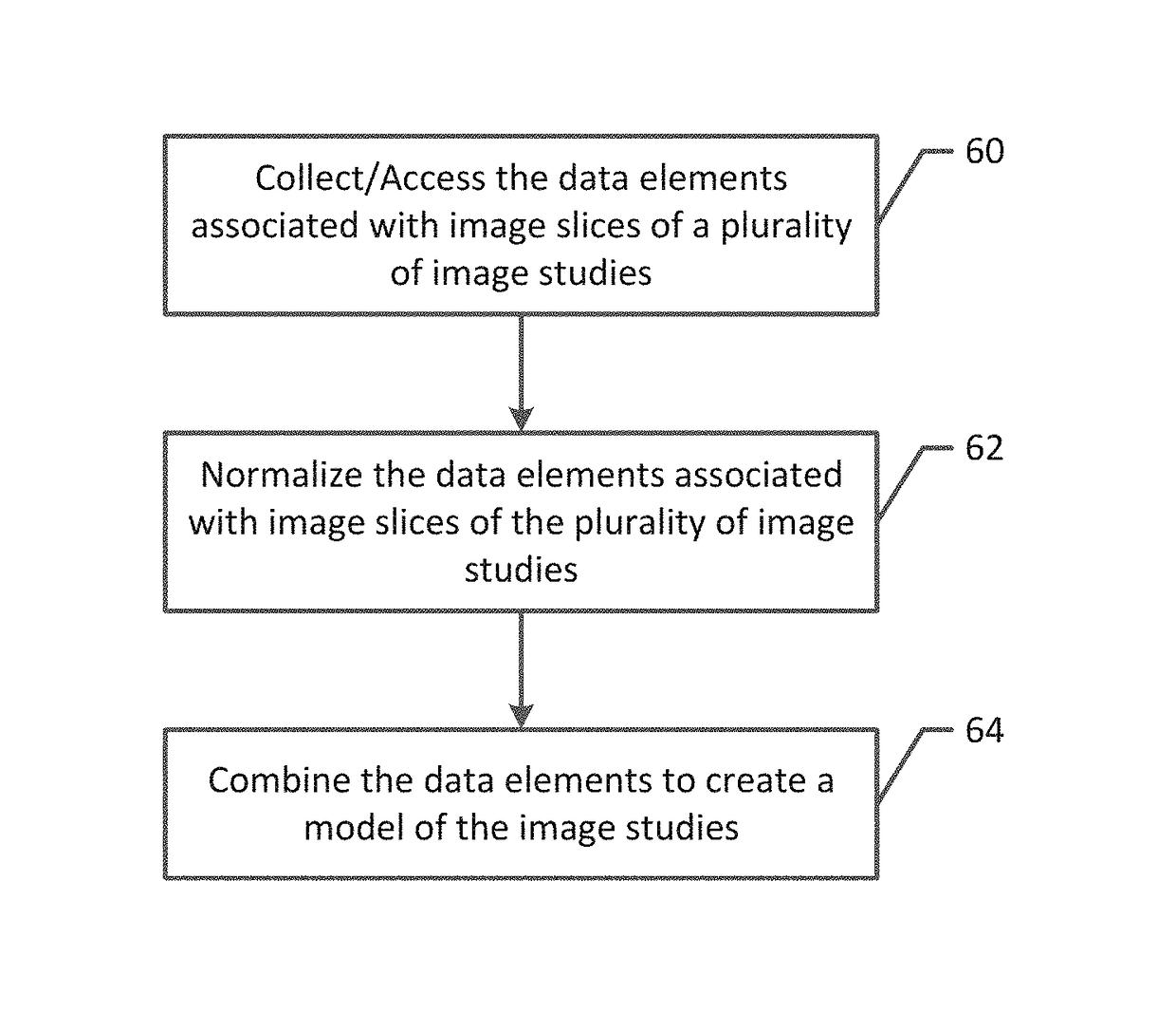
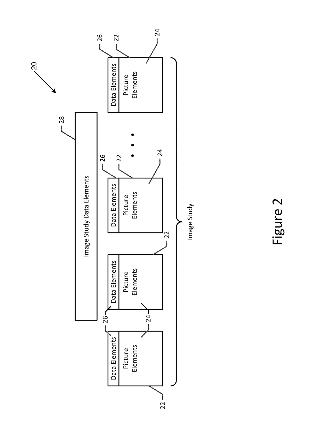
[0020]An image processing system, method and computer program product are provided for detecting a fiducial or an anatomical marker (hereinafter generally referenced as an “anatomical marker”) within an image study. The image processing system, method and computer program product rely, not upon the picture elements (pixels) that comprise the image slices of an image study, but on other data elements that are representative of one or more characteristics of the ...


PUM


Abstract

A method, image processing system and computer program product are provided for detecting an anatomical marker within an image study. In the context of a method, data elements associated with image slices of the image study of a patient are accessed. The data elements are representative of one or more characteristics of the image slices, but do not include picture elements that comprise the image slices. The method also includes reviewing the data elements associated with at least some image slices of the image study of the patient. The method further includes detecting the anatomical marker within the image study based upon a review of the data elements and identifying a subset of the image slices of the image study based upon the anatomical marker or a distance from the anatomical marker.

Description

TECHNOLOGICAL FIELD[0001]An example embodiment relates generally to the review of an image study comprised of a plurality of image slices and, in particular, to the detection of an anatomical marker within an image study in order to permit a subset of the image slices of the image study to be identified based upon the anatomical marker.BACKGROUND[0002]Image studies of a patient may be captured by a plurality of different cross-sectional imaging modalities, such as computed tomography (CT) imaging, magnetic resonance (MR) imaging and the like. The image study may include a plurality of image slices captured at different locations along a patient's body. For example, a patient lying on a table may undergo an image scan with image slices captured at each of a plurality of discrete locations. As a more particular example, the image study of the abdomen of a patient may include a plurality of image slices including image slices of the upper abdomen, images slices of the mid-abdomen and i...

Claims


Application Information

Patent Timeline
Patent Type & Authority Patents(United States)
IPC IPC(8): G06K9/00G06T7/00G16H30/20G16H30/40G16H50/50
CPCG16H50/50G06T2207/10081G06T7/73G06T7/0012G06T2207/10088G16H30/40G16H30/20G06T2207/30204
Inventor NOORDVYK, ALLANREZAEE, MAHMOUD RAMZEPATTI, JAY WALDRON
Owner CHANGE HEALTHCARE HLDG LLC
Why Eureka
Try Eureka