Devices and methods for endovascular access and therapy

a technology for applied in the field of endovascular access and therapy devices and methods, can solve the problems of short response time, achieve the effects of facilitating vascular access, preventing hemorrhage, and facilitating vascular access

Active Publication Date: 2018-11-06
TRAUMATEK SOLUTIONS
View PDF83 Cites 4 Cited by
  • Summary
  • Abstract
  • Description
  • Claims
  • Application Information

AI Technical Summary

Benefits of technology

[0037]In one aspect of the invention, devices and methods provide endovascular therapy, including facilitating establishment of vascular access, placement of endovascular sheaths, catheter tip localization, and administration of vascular occlusion. In one aspect of the current invention, devices and methods are provided for facilitating vascular access and quick, safe, and easy deployment of temporary EAO in order to stop the hemorrhage and enable stabilization of the injured until definitive treatment can be provided. Use of the current invention by trauma teams is expected to result in significant reductions in mortality related to traumatic hemorrhage. Certain elements of the invention may also be utilized alone or in combination for other indications.
[0040]The cannulation device may further include one or more side openings and / or a distal tip, which may be tapered. The cannulation device may also further include a backplate covering the device body at the proximal end of the device body having an opening configured for passing the needle. In other embodiments, the device may further include rollers for pushing the guidewire through the lumen of the device and / or an expandable sheath surrounding the needle. The pressure chamber may be partially pre-filled with biologically acceptable fluid, which may shorten response time.
[0046]Another embodiment of the invention is tip localization device including one or more magnetic field detectors mounted on a support, wherein the device is configured for placement on the outside of a patient's body proximate to the target area of the catheter tip, wherein the one or more magnetic field detectors are capable of detecting the magnetic field emitted by the magnetic catheter tip inside the body of a patient and wherein the one or more magnetic field detectors visually indicate the proximity of the catheter tip. The support may be flexible and the one or more magnetic field detectors may be arranged in an array. The array may include rows and columns of the one or more magnetic field detector which may be configured in arrays including from 3 to 8 rows by from 3 to 8 columns. The distance between the rows and columns of the one or more magnetic field detectors may vary. In one embodiment, the one or more magnetic field detectors include LED which light up when a magnetic field is detected. The flexible support may include a medically acceptable soft pad or cloth. The tip localization device may be configured to remain approximately flat on the patients' body and to prevent extreme overlap between detector fields.
[0048]Yet another embodiment of the invention is a vessel cannulation device having a lumen, wherein the device is configured for automatically inserting a guidewire or sheath in response fluid entering the entering lumen containing fluid at different pressure. Certain embodiments of the device may further include one or more of (1) a pressure sensor such as e.g. a diaphragm, (2) an automatic guidewire release mechanism, (3) a wheel mechanism for pushing a guidewire, (4) a needle and (5) an expandable sheath over the needle. The lumen of the device may be pre-filled with a fluid for shortening response time.
[0057]The vessel cannulation device may also further include a fluid passageway coupled to the pressure sensor. The fluid passageway may be partially or at least partially pre-filled with a biologically acceptable fluid. When the passageways are pre-filled or at least partially pre-filled, the device may be configured to shorten response time. The distal tip of the housing may include needle. The distal tip of the house may also be configured for piercing tissue.
[0082]An alternate embodiment of the invention is a tip localization device including one or more magnetic field detectors mounted on a support, wherein the device is configured for placement on the outside of a patient's body proximate to the target area of the catheter tip, wherein the one or more magnetic field detectors are capable of detecting the magnetic field emitted by the magnetic catheter tip inside the body of a patient and wherein the one or more magnetic field detectors visually indicate the proximity of the catheter tip. The support may be flexible and may include a medically acceptable soft pad or cloth. The one or more magnetic field detectors may be arranged in an array, which may include rows and columns of the one or more magnetic field detectors. For example, the one or more magnetic field detectors may be arranged in an array comprising from 3 to 8 rows by from 3 to 8 columns. The distance between the rows and columns of the one or more magnetic field detectors may vary. The magnetic field detectors may include LED which light up when a magnetic field is detected. The device may be configured to remain approximately flat on the patients' body and to prevent extreme overlap between detector fields.

Problems solved by technology

The pressure chamber may be partially pre-filled with biologically acceptable fluid, which may shorten response time.

Method used


Image

  • Devices and methods for endovascular access and therapy
  • Devices and methods for endovascular access and therapy
  • Devices and methods for endovascular access and therapy

Examples


example 1

nt of Response Time of Vessel Cannulation Device

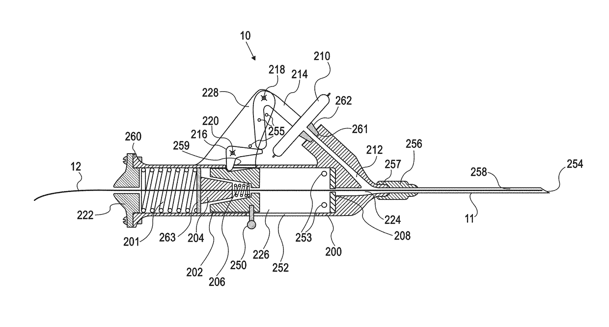
[0375]Tests for measurement of vessel cannulation device response time were performed by puncturing a pressurized elastic tubing system filled with an electrolyte solution, using device embodiment 269 (shown in FIG. 41). The device was connected to a laptop computer via a data acquisition card with sampling input voltage at a rate of 500 Hz and was wired as follows:

[0376]1. Wiring for Identification of Tube Puncture Time:

[0377]One wire was connected to needle 11 and the other wire to a metal tubing connector in fluid communication with the rest of the elastic tubing system. Thus, puncture of the elastic tubing by needle 11 closed an electrical circuit via the electrolyte solution thereby indicating puncture time.

[0378]2. Wiring for Identification of Lever Movement Time:

[0379]Wires were connected to axes 216 and 218 of levers 214 and 220, all of which are made of conductive metals. Before wire advancement, the levers 214 and 220 create ...


PUM


Abstract

The present invention provides for devices and methods for providing endovascular therapy, including facilitating establishment of vascular access, placement of endovascular sheaths, catheter tip localization, and administration of vascular occlusion. The invention includes a vessel cannulation device, an expandable sheath, an occlusion catheter, and a localizer each of which may be provided separately or used as part of a system. One embodiment of the invention is a vessel cannulation device including: a housing having a distal end with a distal tip and a proximal end; a guidewire lumen passing through the housing and at least the distal tip; a sensor coupled to the guidewire lumen; and an advancing member, which is configured for advancing at least one of a guidewire or a sheath and which is operably coupled to the sensor.

Description

CROSS-REFERENCE TO RELATED APPLICATIONS[0001]This application is a continuation-in-part of U.S. application Ser. No. 13 / 708,878 (filed on Dec. 7, 2012), which claims priority to U.S. Provisional Application 61 / 568,111 (filed on Dec. 7, 2011) and U.S. Provisional Application No. 61 / 612,334 (filed on Mar. 18, 2012), the disclosures of which are incorporated by reference in their entireties. This application also claims priority to U.S. Provisional Application No. 61 / 734,925 (filed on Dec. 7, 2012), the disclosure of which is incorporated by reference in its entirety.FIELD OF THE INVENTION[0002]The invention relates to devices and methods for providing endovascular therapy, including facilitating establishment of vascular access, placement of endovascular sheaths, catheter tip localization, and administration of vascular occlusion.BACKGROUND OF THE INVENTIONI. Clinical Need[0003]General:[0004]Vascular access is a crucial element of medical therapy in a vast majority of clinical setting...

Claims


Application Information

Patent Timeline
Patent Type & Authority Patents(United States)
IPC IPC(8): A61M25/01A61M25/06A61B17/12A61M25/09A61M25/00A61M25/10A61B17/00A61B90/00A61B46/10
CPCA61M25/0133A61B17/12031A61B17/12109A61M25/065A61M25/09041A61B17/1214A61M2025/1052A61B17/12168A61B46/10A61B2017/00022A61B2090/064A61M25/0127A61M2025/0024A61B17/12136
Inventor AVNERI, ITZHAKAVNERI, BEN-AMIAVNERI, SHAHARAVNERI, LIOR
Owner TRAUMATEK SOLUTIONS
Why Eureka
Try Eureka