
Cytological image processing device, and method for quantifying characteristics of cytological image

a cytological image and processing device technology, applied in image enhancement, instruments, optical elements, etc., can solve problems such as diagnosis accuracy, and achieve the effect of improving diagnosis accuracy and reducing disagreemen

Active Publication Date: 2019-12-03
AMCAD BIOMED CORP
View PDF5 Cites 0 Cited by
  • Summary
  • Abstract
  • Description
  • Claims
  • Application Information

AI Technical Summary

Benefits of technology

The present invention provides a way to analyze and segment cytological images, which can be useful in diagnosing patient illness. By dividing the image into a nuclear and cytoplasmic part and tracking nuclear blocks, relevant parameters can be measured and summarized in an accessible way. This can help doctors identify specific features of the image and make more accurate diagnoses. This invention aims to reduce diagnostic inconsistencies and improve the accuracy of medical diagnosis.

Problems solved by technology

However, in the case where the diagnosis is made only depending on the cytological image without performing advanced processing on the cytological image to obtain relevant quantified parameters or analyzed images, different doctors may give quite different diagnosis results for the same cytological image, and the excessively subjective diagnosis results will influence the accuracy of diagnosis when determining the illness condition of the patient.

Method used


Image

  • Cytological image processing device, and method for quantifying characteristics of cytological image
  • Cytological image processing device, and method for quantifying characteristics of cytological image
  • Cytological image processing device, and method for quantifying characteristics of cytological image

Examples


first embodiment

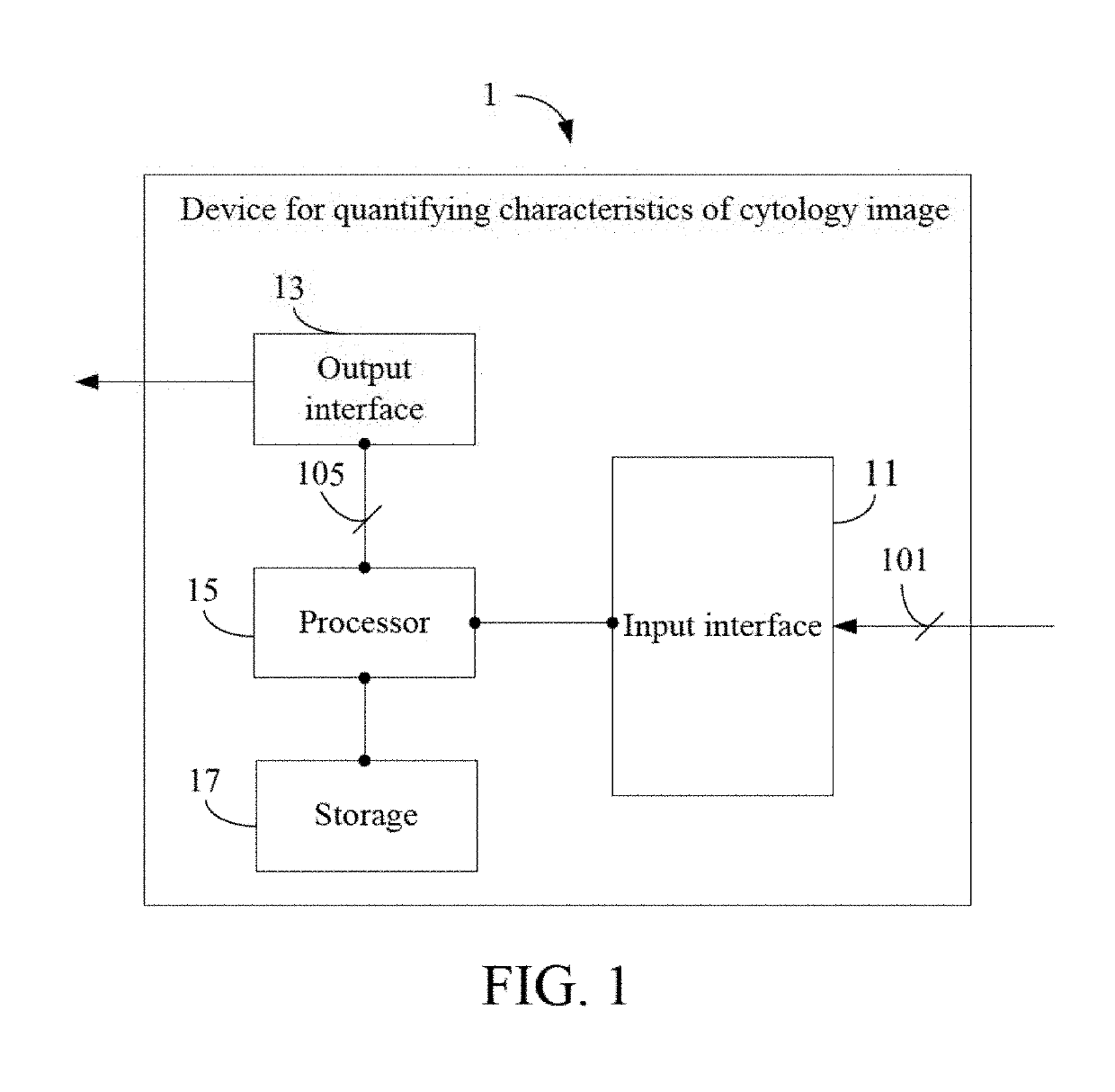
[0030]the present invention is as shown in FIG. 1 and FIG. 2A. FIG. 1 is a schematic view of a cytological image processing device 1 according to the present invention. The cytological image processing device 1 comprises an input interface 11, an output interface 13, a processor 15 and a storage 17. The processor 15 is electrically connected to the input interface 11, the output interface 13 and the storage 17.

[0031]The input interface 11 is used to receive a cytological image 101 which has been stained (e.g., a cytological image with a magnification factor of 400×, as shown in FIG. 7A, including a background part 701, nuclei 703, and cytoplasm 705) generated by a light microscope (or any image sensor having a high magnification factor). The input interface 11 may be a wired interface (e.g., a USB transmission interface, but not limited thereto) or a wireless transmission interface (e.g., a WIFI transmission interface), and it may be connected to the light microscope or connected to...

second embodiment

[0102]The edge pixels of the line segments which are not eliminated after the above operations are edge pixels of a normal nucleus, so the processor 15 can obtain an edge matrix and connection information of the edge pixels of the edge matrix. In this way, the processor 15 can incorporate the edge matrix obtained based on the temporary background-subtracted images 103_temp and the connection information of the edge matrix into the integrated edge matrix generated in the second embodiment and the integrated connection information of the edge pixels of the integrated edge matrix respectively through the above operations, thereby segmenting the nuclear block 1101 in the background-subtracted image 103 as accurately as possible.

[0103]In addition, in the above embodiments, after segmenting the nuclear blocks, for the cytoplasmic part, the processor 15 can further determine whether the cytoplasmic part has a plurality of pixels which are located within the segmented nuclear blocks and are...


PUM

PropertyMeasurementUnit
coloraaaaaaaaaa
color ratioaaaaaaaaaa
weight proportionaaaaaaaaaa

Abstract

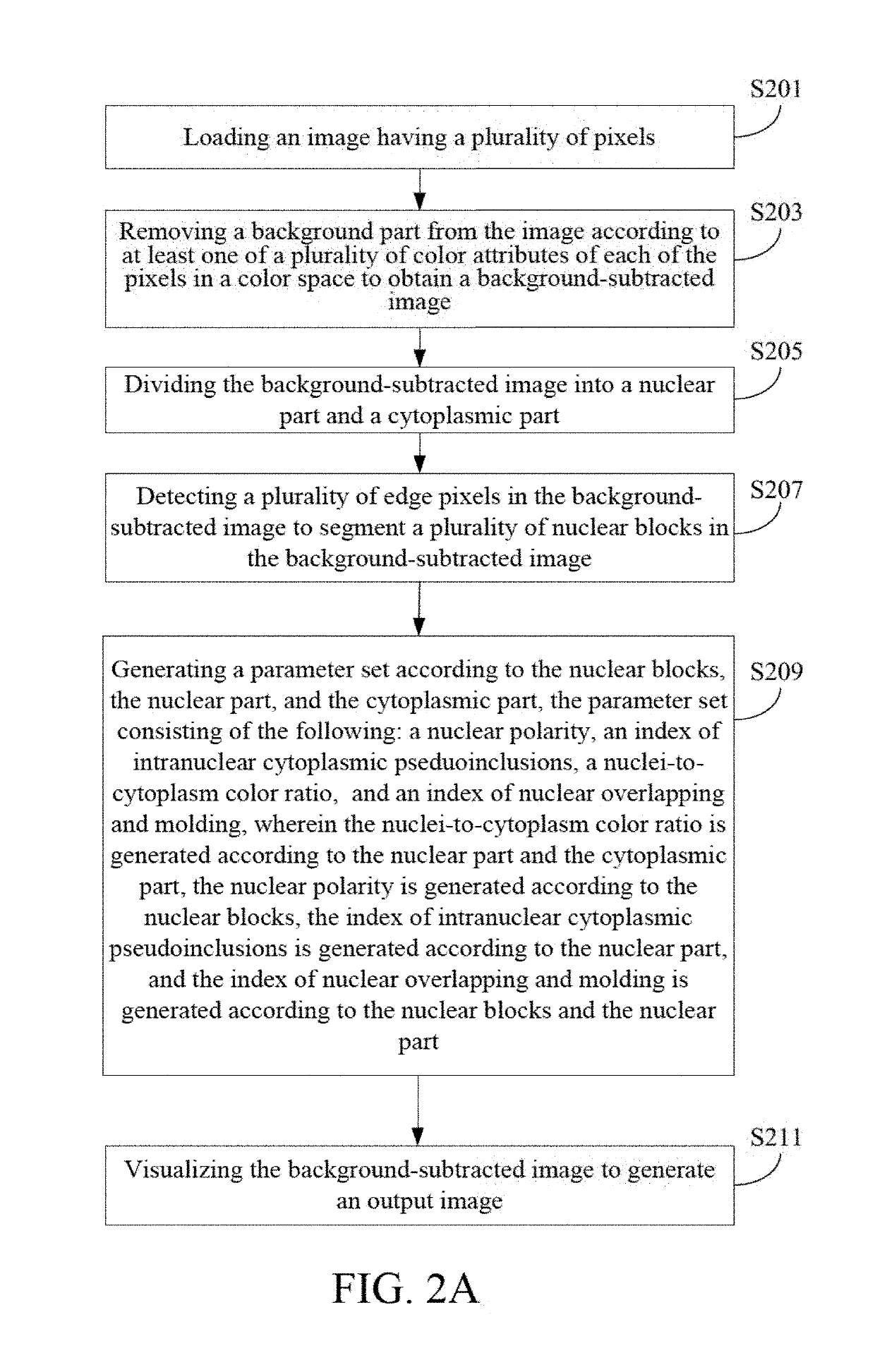
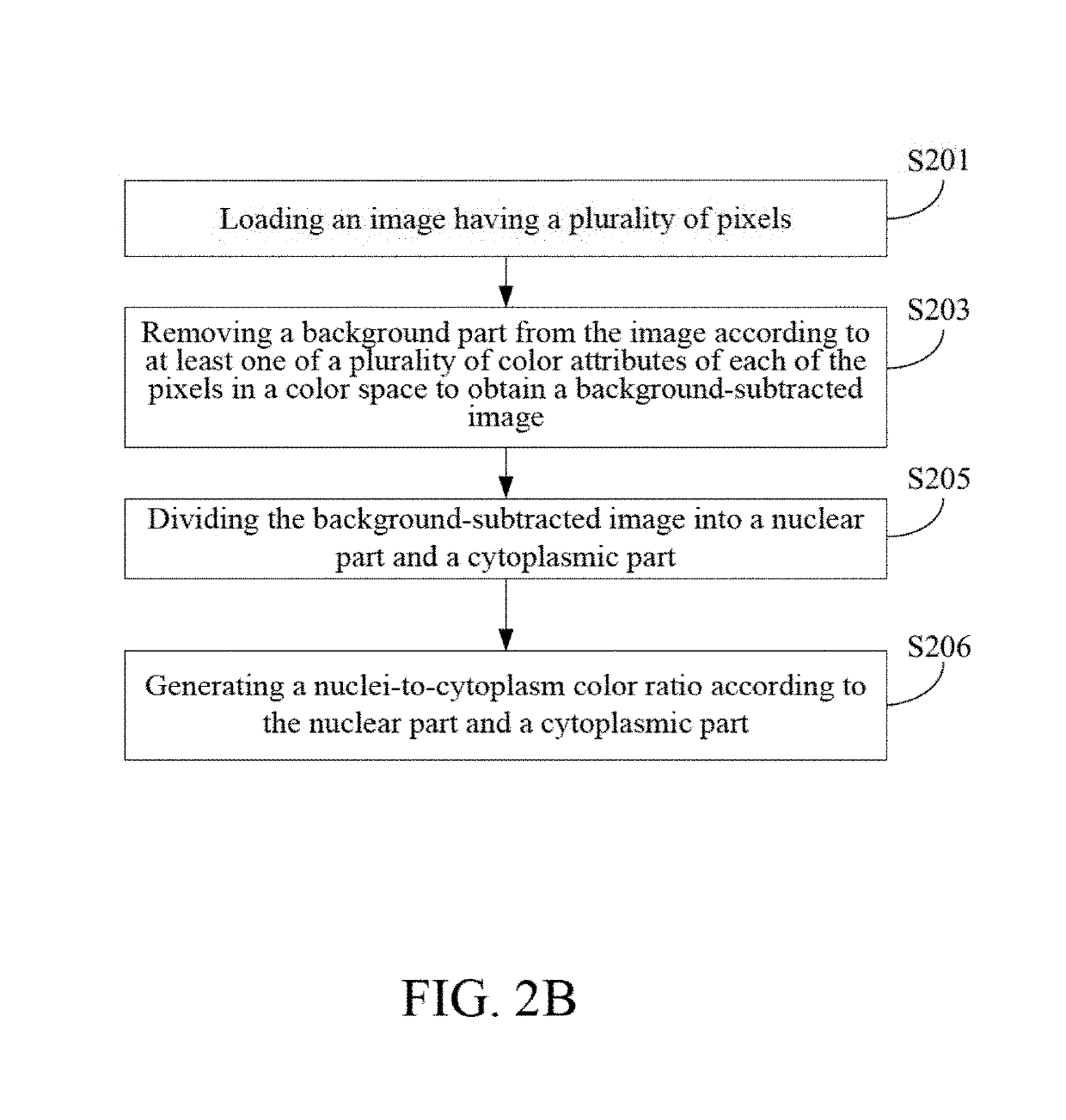
A cytological image processing device, a method for quantifying characteristics of cytological image and a non-transitory computer readable medium thereof are provided. The cytological image processing device executes the method including the steps: loading a cytological image with a plurality of pixels; removing a background part from the cytological image according to at least one of a plurality of color attributes of each pixel of the cytological image in a color space to obtain a background-subtracted image; dividing the background-subtracted image into a nuclear part and a cytoplasmic part; detecting a plurality of edge pixels in the background-subtracted image to segment a plurality of nuclear blocks in the background-subtracted image; generating a parameter set according to the nuclear blocks; and generating an output image by visualizing the background-subtracted image.

Description

[0001]This application claims the benefit of and is a non-provisional of co-pending U.S. Provisional Application Ser. No. 62 / 419,928 filed on Nov. 9, 2016, which is hereby expressly incorporated by reference in its entirety for all purposes.CROSS-REFERENCES TO RELATED APPLICATIONS[0002]Not applicable.BACKGROUND OF THE INVENTIONField of the Invention[0003]The present invention relates to a cytological image processing device, and a method for quantifying characteristics of cytological image. More particularly, the cytological image processing device of the present invention executes the method for quantifying characteristics of cytological image to obtain nuclear blocks in a cytological image, and outputs a parameter set and a visualized image according to the nuclear blocks.Descriptions of the Related Art[0004]With the rapid development of the science and technology, technologies for digitalizing data and images have gradually been mature. Electronic products (e.g., computers, table...

Claims


Application Information

Patent Timeline
Patent Type & Authority Patents(United States)
IPC IPC(8): G06T7/00G06T7/90G02B21/36G06T7/194G06K9/00G02B21/06G06T7/11G06K9/46G06T7/136G06T7/12G01N1/30G06K9/62G06T7/181G06T7/73G06V10/44G16B45/00
CPCG06K9/6202G06T7/73G06T7/194G02B21/06G06K9/4604G06T7/0012G06T7/0008G06T7/11G06T7/181G06T7/12G01N1/30G06T7/136G06K9/0014G06K9/00127G06K9/6232G02B21/365G06T7/90G06T2207/10056G06T2207/20132G06T2207/30024G06T2207/10024G06V20/69G06V20/695G06V10/44
Inventor CHEN, ARGONHSIAO, YI-HSIENCHANG, TIEN-CHUNJAN, I-SHIOWSHIH, SHYANG-RONGWANG, HSUAN-MAO
Owner AMCAD BIOMED CORP
Learn More