
Method and apparatus for generating a multi-slice data set of a heart

a multi-slice data and heart technology, applied in the field of three-dimensional multi-slice data sets of hearts, can solve the problems of infrequent use of methods and inability to reliably calculate heart functions, and achieve the effect of improving the resolution of three-dimensional recordings

Active Publication Date: 2020-01-07
SIEMENS HEALTHCARE GMBH
View PDF4 Cites 0 Cited by
  • Summary
  • Abstract
  • Description
  • Claims
  • Application Information

AI Technical Summary

Benefits of technology

[0007]An object of the present invention is to improve the resolution of three-dimensional recordings (data set acquisitions) of the heart in patients whose heartbeat intervals are arrhythmic.
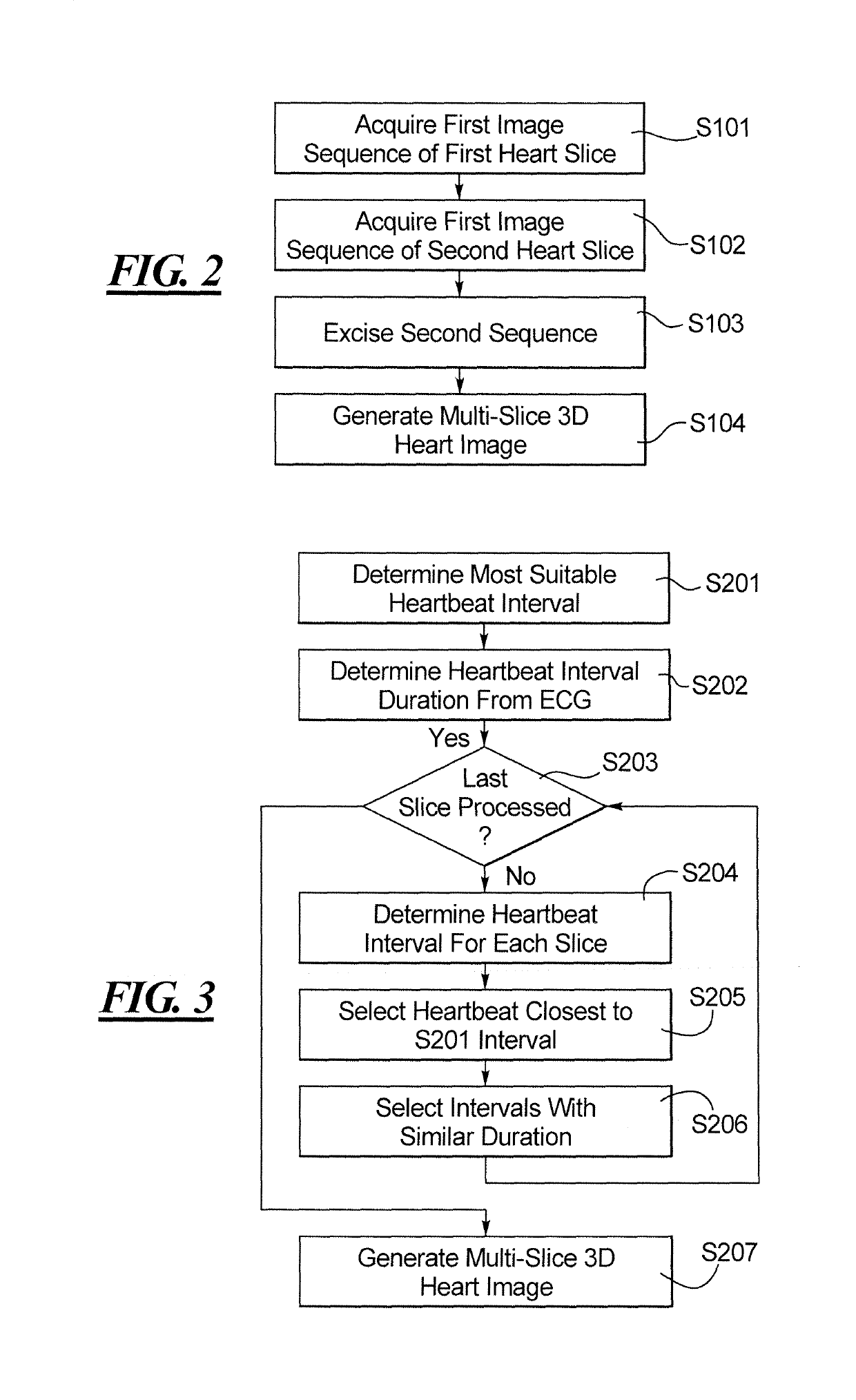
[0008]According to a first aspect of the invention, this object is achieved by a method for generating a three-dimensional multi-slice data set of a heart, having the following steps. A first image sequence is acquired that includes several two-dimensional images in a first slice of the heart during a duration of at least one heartbeat interval. A second image sequence is acquired that includes a number of two-dimensional images in a second slice of the heart during the duration of a number of heartbeat intervals. The second image sequence is excised by selecting therefrom the two-dimensional images that lie in a heartbeat interval that has the duration closest to the duration of the heartbeat interval of the first slice. A three-dimensional multi-slice data set is generated by combining the first image sequence and the excised second image sequence. When this method is applied to multiple slices, the volume of the heart can be reconstructed over a large scale. As a result, the advantage is achieved that heartbeats with similar heartbeat intervals are selected for the different slices and an arrhythmia of the heartbeat can be balanced out. An automatic functional calculation of the heart helps physicians to better assess the position of patients who are suffering heart arrhythmia.
[0009]In an embodiment of the method, the method includes the step of determining the duration of one of the heartbeat intervals by recording an electrocardiogram during the acquisition of the first image sequence and / or the second image sequence. As a result, the advantage is achieved such that the duration of the heartbeat interval can be determined easily.

Problems solved by technology

The other basic method is prospective gating, which is used for patients whose heartbeat frequency changes during the acquisition duration, since the changes in the interval duration can generate artifacts during retrospective gating.
Although prospective gating is the sole solution for patients with arrhythmic heartbeats, this method is infrequently used because of its lower resolution compared with retrospective gating.
A further factor with prospective gating is, however, that the heart function cannot be reliably calculated with the acquisition of a single heartbeat, since the heartbeat has a different duration for each recorded individual slice of the heartbeat, which results in different function results.

Method used


Image

  • Method and apparatus for generating a multi-slice data set of a heart
  • Method and apparatus for generating a multi-slice data set of a heart
  • Method and apparatus for generating a multi-slice data set of a heart

Examples


Embodiment Construction

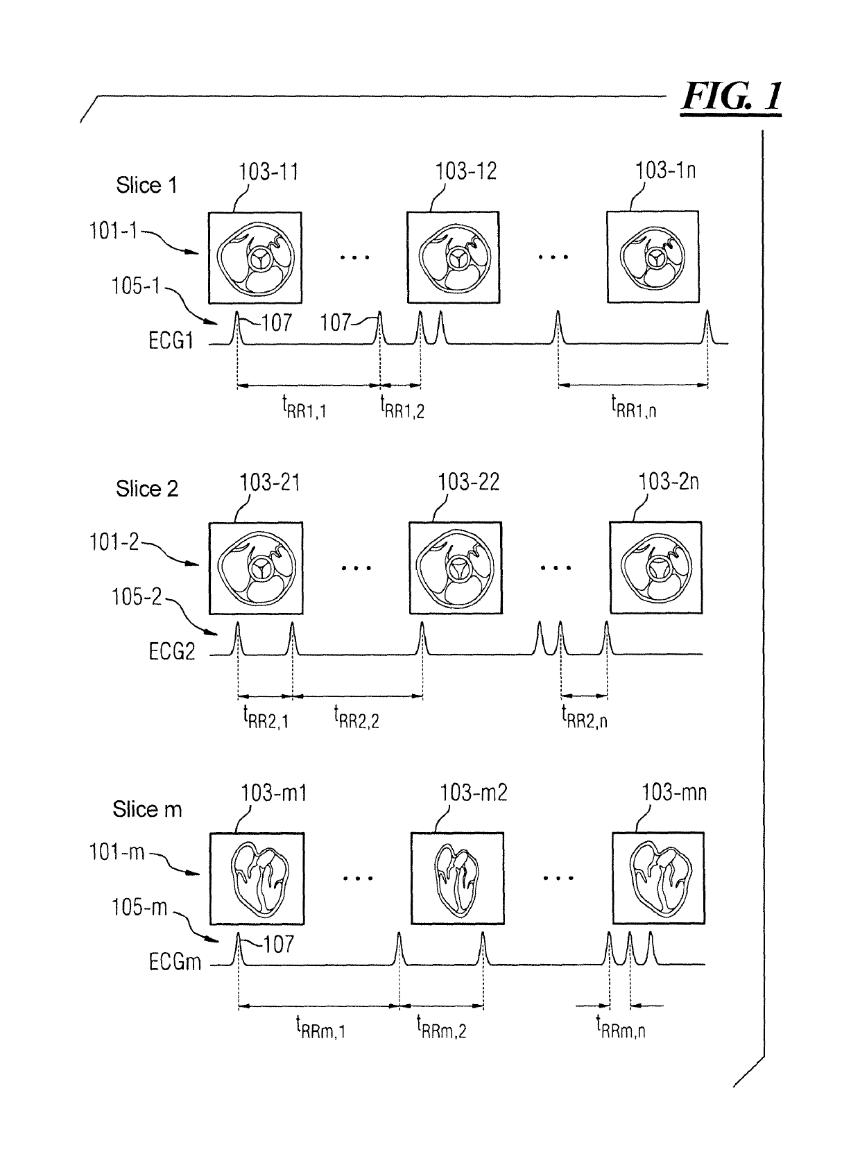
[0028]FIG. 1 shows a representation of image sequences 101-1, . . . , 101-m in different slices of the heart. The image sequences 101-1, . . . , 101-m are generated by means of a tomography method, which can determine the inner spatial structure of an object and represent the same in the form of sectional images.

[0029]FIG. 1 represents the situation when acquiring MR realtime data in a number of slices using prospective gating which is obtained over several heartbeats. For each slice orientation, like for instance the short axis (SAX) or the long axis (LAX), a number of heartbeats are plotted and stored together with an electrocardiogram 105-1, . . . , 105-m. The duration trr(m, n) is the duration of the heartbeat interval (RR-interval) of the slice m for the heartbeat n.

[0030]Each of the image sequences 101-1, . . . , 101-m includes a number of two-dimensional images 103-11, . . . , 103-mn, which represent a sectional image of the heart in a certain slice. The image sequence 101-1 ...


PUM


Abstract

In a method and apparatus for generating a three-dimensional multi-slice data set of a heart, a first image sequence with a number of two-dimensional images is acquired in a first slice of the heart during a duration of at least one heartbeat interval, and a second image sequence with a number of two-dimensional images is acquired in a second slice of the heart during the duration of a number of heartbeat intervals. The second image sequence is excised by selecting the two-dimensional images thereof that lie in a heartbeat interval closest to the duration of the heartbeat interval in the first slice. The three-dimensional multi-slice data set is generated by combining the first image sequence and the excised second image sequence.

Description

BACKGROUND OF THE INVENTION[0001]Field of the Invention[0002]The present invention concerns a method for generating a three-dimensional multi-slice data set of a heart, and a tomography apparatus for generating a three-dimensional multi-slice data set of a heart.[0003]Description of the Prior Art[0004]Two methods of acquiring moving images of a heart and calculating a heart function therefrom currently exist in cardiac magnetic resonance tomography (MRI—Magnetic Resonance Imaging). With retrospective gating, the heartbeat frequency of the patient barely changes during the measurement time, so that a reconstruction of an average heartbeat cycle is enabled. This average heartbeat cycle effectively reproduces the typical heartbeat of the patient. The other basic method is prospective gating, which is used for patients whose heartbeat frequency changes during the acquisition duration, since the changes in the interval duration can generate artifacts during retrospective gating. Although...

Claims


Application Information

Patent Timeline
Patent Type & Authority Patents(United States)
IPC IPC(8): A61B6/03G06T15/08G06T7/20G06T11/00A61B6/00A61B8/13A61B8/08A61B5/352
CPCA61B6/03A61B6/503G06T11/008G06T7/20G06T15/08G06T2207/10072A61B8/0883G06T2207/20182A61B8/13G06T2211/40G06T2207/10016G06T2207/20221G06T2207/30048A61B6/032A61B6/486A61B6/5235A61B6/5288G06T2211/412
Inventor DE OLIVEIRA, ANDRE
Owner SIEMENS HEALTHCARE GMBH
Learn More