System and method for automated funduscopic image analysis

a funduscopic and image analysis technology, applied in the field of automatic funduscopic image analysis, can solve the problems of inability to demonstrate the reliability and accuracy of a trained human, no symptoms a patient can detect, and high cost of manual processes, so as to improve network performance, simplify the problem, and increase the degree of freedom of input data set.

Active Publication Date: 2020-07-21
RETINOPATHY ANSWER LTD
View PDF41 Cites 4 Cited by
  • Summary
  • Abstract
  • Description
  • Claims
  • Application Information

AI Technical Summary

Benefits of technology

[0089]The training data employ may be the entire retinal image with regional classifications associated as labels, though this increases the degrees of freedom of the input data set, which may complicate the problem without corresponding improvement of network performance. That is, retinopathy in one region of the retina is very similar to retinopathy in other regions, and therefore the training need not be region-specific. Of course, there may indeed be region-specificity, and the present technology does not ignore or disclaim this, and rather finds that adequate performance is achievable while making the region-independent presumption. For example, in a 1000×1000 input image, there are 106 pixels, and without simplification, a NN would have to have at least 106 input neurons. On the other hand, 64×64 pixel regions provide sufficient precision to identify pathology, requiring at least 4096 input neurons, about 250× fewer. Likewise, the region-independent presumption may permit reduction in at least an entire hidden layer of the network which is designed to process regional differences.
[0090]Prior to processing, the image may be normalized with respect to color, brightness, etc., in a deterministic statistical or rule-based process. While this normalization may be achieved within the NN, this can also be achieved independent of the network, leading to a simpler implementation of the network.
[0091]In some cases, the regions may be normalized after segmentation, or as part of the segmentation process. This permits segmentation to be based on a statistical or rule based process that establishes regional boundaries on a basis other than shape or space. Again, normalization preceding the NN simplifies the NN implementation, but also relieves the NN of learning the pre-normalized features. By employing expert determination of features that may be extracted from the image by a normalization process, the specificity of the NN for the features which are not normalized is increased.
[0101]The precise balance needed between type I and II errors, as outlined above, will depend on the intended use of the system. For a hospital setting minimizing type II error may be the most important, while for general screening via opticians it might be most important to minimize type I error and avoid causing needless worry to customers.
[0103]The algorithm was found to be highly effective at marking regions of the retinal image with indications of disease, demonstrating that the image processing / region matching technology was effective and efficient.

Problems solved by technology

There are many types of retinopathy which can be detected on the image, from hemorrhages to tumors, and although many such diseases can lead to serious consequences (including blindness), the early stages usually cause no symptoms a patient can detect.
This manual process is costly, but to date is the most reliable way to process these images.
Despite years of research, no automated system has yet been able to demonstrate the levels of reliability and accuracy a trained human can achieve.
Most interpretations of medical images are performed by physicians; however, image interpretation by humans is limited due to its subjectivity, large variations across interpreters, and fatigue.
Human examiners may be inconsistent in their interpretations, and prone to error.
Therefore, even obtaining training data is not without difficulty.
First, CNNs require a large amount of labeled training data, a requirement that may be difficult to meet in the medical domain where expert annotation is expensive and the diseases (e.g., lesions) are scarce.
Second, training a DCNN requires large computational and memory resources, without which, the training process would be extremely time-consuming.
Third, training a DCNN is often complicated by overfitting and convergence issues, which often require repetitive adjustments in the architecture or learning parameters of the network to ensure that all layers are learning with comparable speed.
However, empirical adjustments to the process characterize the state of the art, with significant differences in approach, implementation, and results are observed in the art even when using similar algorithms for similar goals.
Note that the training of a network cannot be updated, and any change in the algorithm or training data requires re-optimization of the entire network.
Unsupervised representation learning methods such as Restricted Boltzmann Machines (RBM) may outperform standard filter banks because they learn a feature description directly from the training data.
The RBM is trained with a generative learning objective; this enables the network to learn representations from unlabeled data, but does not necessarily produce features that are optimal for classification.
The lack of publicly available ground-truth data, and the difficulty in collecting such data per medical task, both cost-wise as well as time-wise, is a prohibitively limiting factor in the medical domain.
The fact that expert tasks are being outsourced to non-expert users may lead to noisy annotations introducing disagreement between users.
Likewise, use of uncontrolled clinical outcome data, or user data as a basis for feedback, may lead to poor outcomes, due to lack of standardization and control.
Many issues arise in combining the knowledge of medical experts with non-professionals, such as how to combine the information sources, how to assess and incorporate the inputs weighted by their prior-proved accuracy in performance and more.
This perhaps means that the results do not capture and exploit the skill, knowledge and insight of the experts.

Method used


Image

  • System and method for automated funduscopic image analysis
  • System and method for automated funduscopic image analysis
  • System and method for automated funduscopic image analysis

Examples


Embodiment Construction

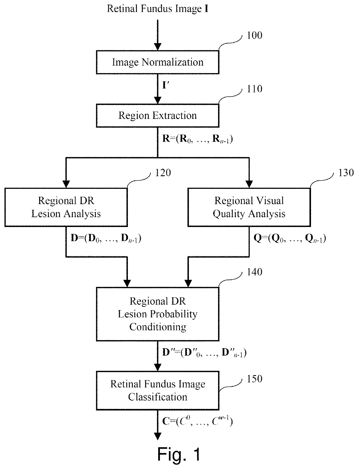
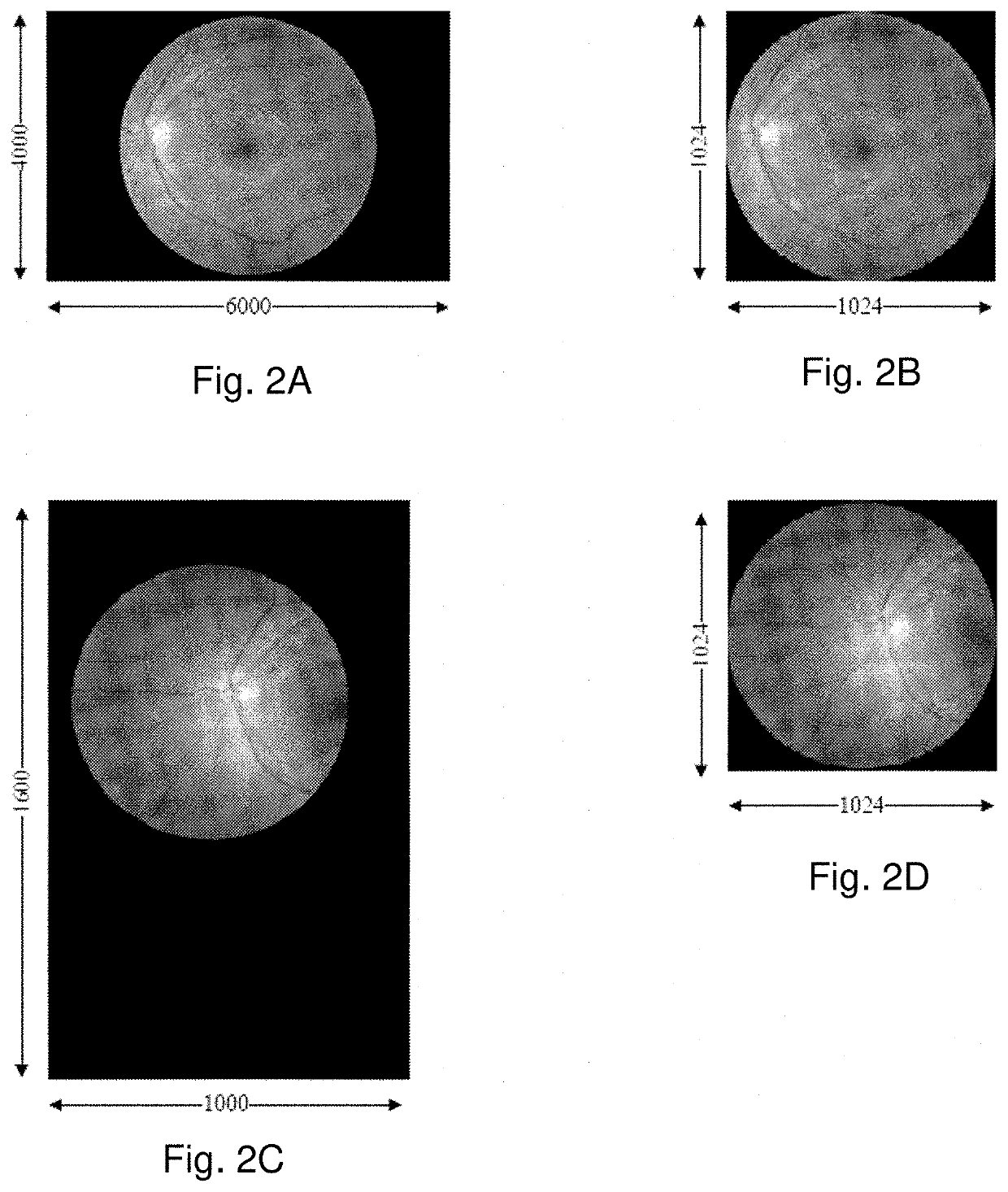
[0126]FIG. 1 illustrates an embodiment of the invention. Given a retinal fundus image I, block 100 performs image normalization to produce a normalized image I′. This normalization step involves identifying the retinal disc in I and resizing it to a fixed size, i.e., to a diameter of p pixels in an image of p×p pixels, where, typically, p=1024. This is illustrated in FIGS. 2A-2D, where FIGS. 2A and 2C show pre-normalized retinal fundus images I and FIGS. 2B and 2D show the corresponding normalized retinal fundus images I′. Automatic detection of the retinal disc is a straightforward task which entails the detection of a relatively bright disc on a relatively dark and uniform background. As such, a number of well-known image processing techniques may be used, such as edge detection, circular Hough transform, etc. More details of such techniques may be found in Russ, John C. The image processing handbook. CRC press, 2016.

[0127]Next, in block 110 of FIG. 1, retinal regions are extracte...


PUM


Abstract

A system and method of classifying images of pathology. An image is received, normalized, and segmented normalizing the image; into a plurality of regions; A disease vector is automatically determining for the plurality of regions with at least one classifier comprising a neural network. Each of the respective plurality of regions is automatically annotated, based on the determined disease vectors. The received image is automatically graded based on at least the annotations. The neural network is trained based on at least an expert annotation of respective regions of images, according to at least one objective classification criterion. The images may be eye images, vascular images, or funduscopic images. The disease may be a vascular disease, vasculopathy, or diabetic retinopathy, for example.

Description

CROSS REFERENCE TO RELATED APPLICATION[0001]The present application claims priority from U.S. Provisional Patent Application No. 62 / 490,977, filed Apr. 27, 2017, the entirety of which is expressly incorporated herein by reference.FIELD OF THE INVENTION[0002]The present invention relates to the field of automated funduscopic image analysis using neural networks (“NN”) or deep neural networks (“DNN”).BACKGROUND OF THE INVENTION[0003]Each of the references cited herein are expressly incorporated herein by reference in their entirety.[0004]The human retina is readily photographed using a variety of commonly available specialist cameras. In the UK most opticians, in particular the major chains, have such cameras in their practices already and a retinal photograph is increasingly offered as part of a standard eye test.[0005]Retinal images may be used to assess many eye diseases—or in general retinopathy—and over recent years as the resolution of digital cameras have increased, has become ...

Claims


Application Information

Patent Timeline
Patent Type & Authority Patents(United States)
IPC IPC(8): G06T7/00A61B3/14A61B3/00A61B3/12G06K9/46G06N3/08G06K9/62G06T7/11G06K9/42G06K9/00
CPCG06K9/42G06T7/0016G06T7/11G06K9/4671A61B3/12G06K9/6257G06K9/0061A61B3/14G06K9/00617G06N3/08G06K9/6269A61B3/145A61B3/0025G06T2207/20081G01N2800/16G06K2209/05G06T2207/20084G06T2207/30041G06V40/197G06V10/462G06V2201/03G06V40/193G06F18/2148G06F18/2411
Inventor PASCHALAKIS, STAVROSBOBER, MIROSLAW
Owner RETINOPATHY ANSWER LTD
Why Eureka
Try Eureka