Proteasome modulation for treatment of corneal disorders

a technology of proteasomes and corneal disorders, applied in the field of proteasome modulation for the treatment of corneal disorders, can solve the problems of increasing the activity of cpr, and achieve the effects of reducing corneal transparency, increasing globlet cells, and increasing conjunctival proteins

Active Publication Date: 2020-08-11
LOS ANGELES BIOMEDICAL RES INST AT HARBOR UCLA MEDICAL CENT
View PDF4 Cites 0 Cited by
  • Summary
  • Abstract
  • Description
  • Claims
  • Application Information

AI Technical Summary

Benefits of technology

[0033]Advantages of this approach are numerous. One important advantage of the invention is the improvement in vision of patients at risk for or suffering from corneal opacification. Another advantage is that the invention provides for a non-invasive alternative to treatment of corneal opacification.
[0034]Terms used in the claims and specification are defined as set forth below unless otherwise specified.
[0035]The term “proteasome modulator” refers to any chemical or drug composition or substance that leads to altered proteasome function, including altered proteasome activity or proteasome abundance in the subject.
[0036]The term “corneal opacification” includes conjunctivalization, corneal scarring and corneal conditions resulting in corneal haze, reduction in corneal transparency, increased globlet cells in the cornea, increased conjunctival proteins in the cornea, increased Muc5ac protein in the cornea or increased keratin proteins in the cornea.
[0037]The term “treatment” refers to a medical method for attempting to cause an improvement in the condition of a disease state, including prophylaxis, lessening in the severity or progression, remission, or cure thereof.
[0038]The term “in vivo” refers to processes that occur in a living organism.

Problems solved by technology

In an embodiment, the method results in increased activity of the CPR more than 72 hours after treatment.

Method used


Image

  • Proteasome modulation for treatment of corneal disorders
  • Proteasome modulation for treatment of corneal disorders
  • Proteasome modulation for treatment of corneal disorders

Examples


example 1

ced Model of Opacification

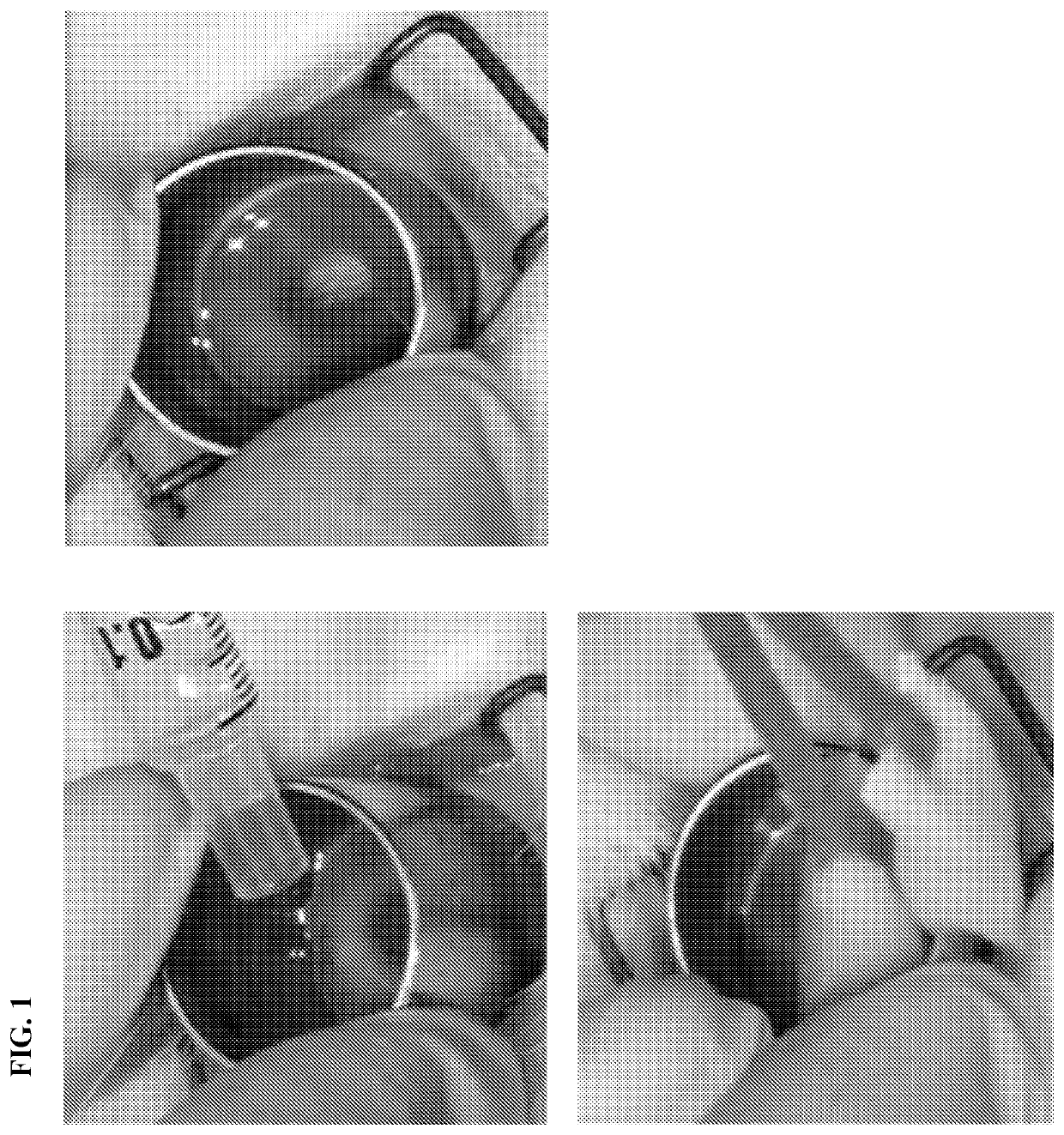
[0071]Our model of LSCD-induced opacification achieved total removal of limbal stem cells and obtained stable LSCD. The experimental protocol was performed according to the following schedule: 1) administration of anesthesia, 2) perform lamellar limbectomy to create LSCD, 3) follow up for 3 months until the pathology is stable, 4) administer PS-341 treatment by topical administration (FIG. 1) of the pre-determined optimal dose of PS-341 every 72 hours for one month. Based on our in vitro study, one drop of PS-341 optimal dose was administered every 72 hours for one month. Corneas were routinely examined in the scheduled follow ups. 5) Grading of corneal opacification was performed based on the visibility of the iris details and scored as: Grade 0: clear cornea with visible iris details; Grade 1: mild opacification with less distinct iris details; Grade 2: moderate opacification if the iris is poorly visible; Grade 3: severe opacification if the iris is not ...

example 2

al Cornea is Covered with Keratin 13 and Keratin 4 in a LSCD-Induced Model of Opacification

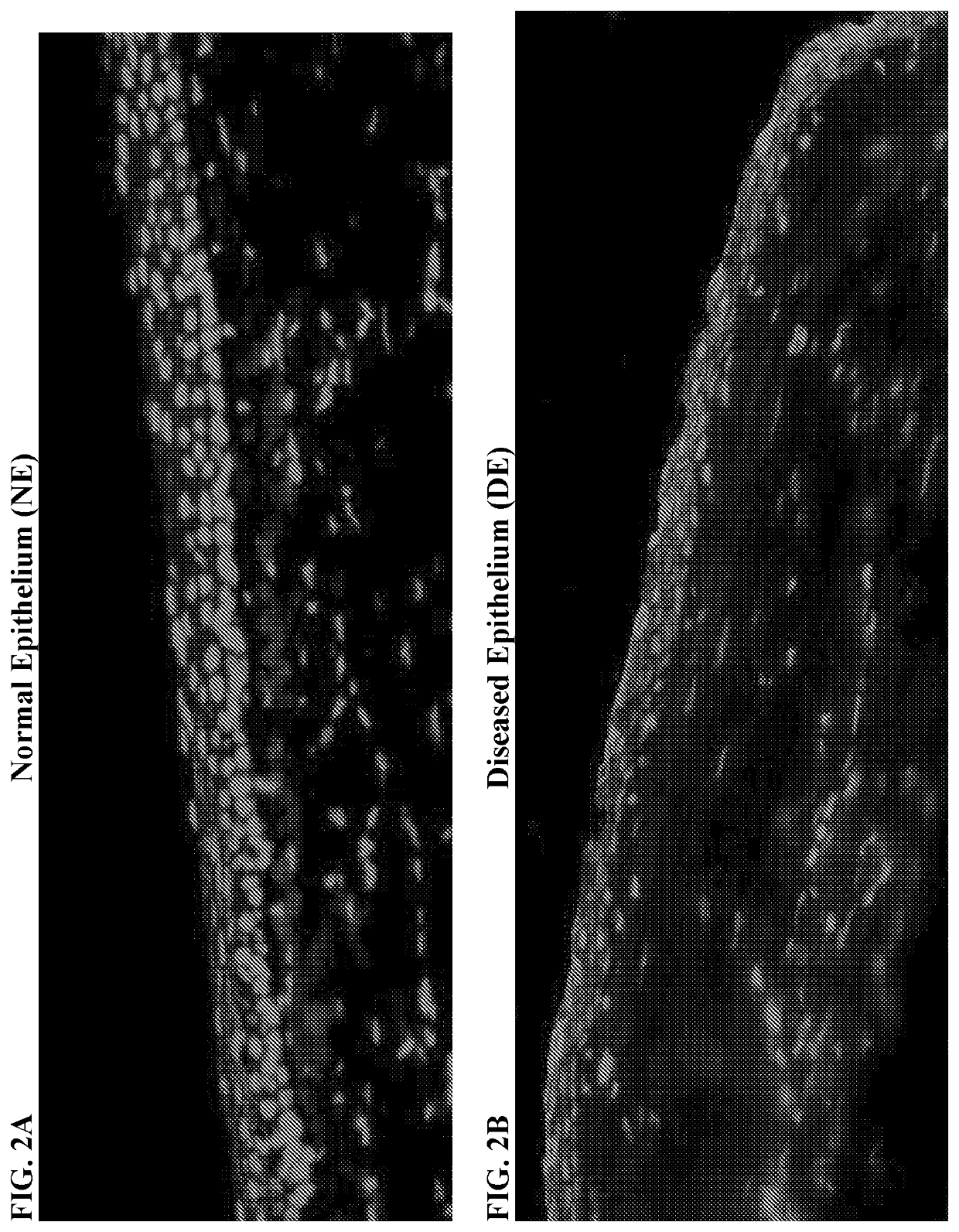
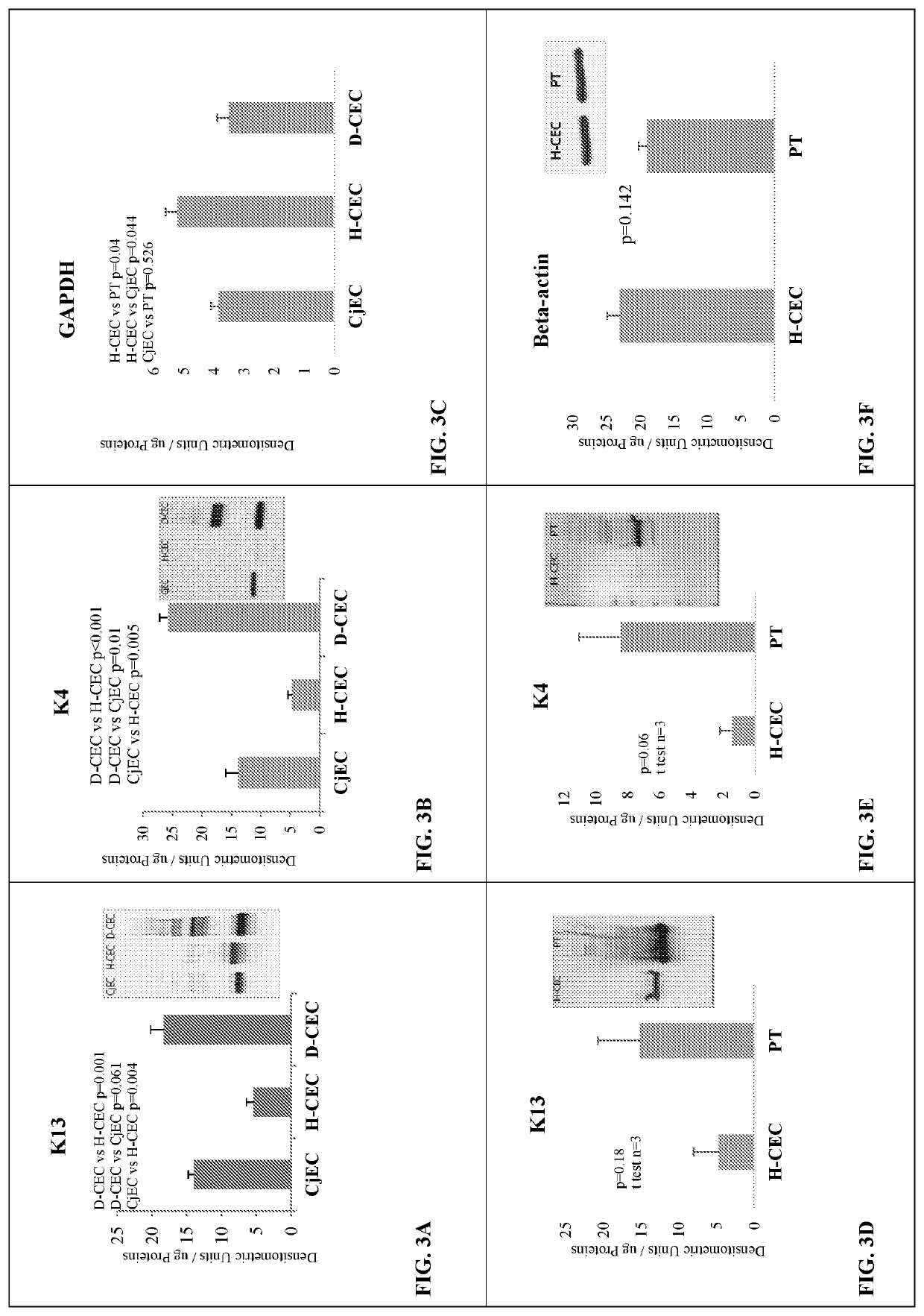
[0072]Results from histopathological examination of the LSCD-induced opacification model showed that while normal cornea stained K13 positive only on the conjunctival side, K13 markedly covered central cornea of LSCD diseased cornea, as shown by the cytoplasmic cellular staining on the top of the tissue sections in FIGS. 2A and 2B, respectively. K4 was also detected in the conjunctiva-limbal area and central cornea of LSCD diseased cornea (data not shown). In addition, pannus tissue harvested from the LSCD diseased cornea was found positive for K4 (data not shown). The results show that the central cornea is covered with K13 and K4 in corneas from animals with LSCD and not in normal cornea. There was K13 and K4 accumulation with additional higher molecular weight bands in diseased corneal epithelial cells (D-CEC) compared to healthy corneal epithelial cells (H-CEC) indicating K13 and K4 posttr...

example 3

asome Activity is Decreased in LSCD Corneal Epithelium

[0073]Experiments made on collected corneal epithelial cells from LSCD rabbits showed that all CPR activities were significantly decreased in corneal diseased epithelium (DE) (FIG. 4).


PUM

PropertyMeasurementUnit
diameteraaaaaaaaaa
diameteraaaaaaaaaa
diameteraaaaaaaaaa

Abstract

Disclosed herein are methods and compositions useful for preventing or reducing corneal haze of opacification resulting from Limbal Stem Cell Deficiency (LSCD). The invention comprises a method of preventing or treating corneal opacification, comprising administering to a subject a sufficient amount of a proteasome modulator. The invention also comprises a method of preventing or treating corneal opacification, comprising administering to a subject a sufficient amount of a proteasome modulator. In addition, the invention comprises a method of administering to a subject suffering from corneal opacification with a sufficient amount of proteasome modulator, resulting in reduction of Keratin proteins in the cornea of the subject.

Description

CROSS REFERENCE TO RELATED APPLICATIONS[0001]This application is a national stage of International Application No. PCT / US2016 / 061656 filed Nov. 11, 2016, which claims the benefit of U.S. Provisional Application No. 62 / 255,338, filed Nov. 13, 2015, the disclosure of which is hereby incorporated by reference.BACKGROUND OF THE INVENTIONField of the Invention[0002]Corneal opacification resulting from Limbal Stem Cell Deficiency (LSCD) leads to abnormal accumulation of protein aggregates in the cornea. Keratins are a group of proteins that form intermediate filaments in epithelial cells. They are divided in two groups: type I Keratins (small, acidic, including K9-K20) and type II Keratins (large, neutral to basic, including K1-K8). They are usually found in pairs that include a type I and type II and are highly tissue-specific. Corneal epithelial cells are characterized by the expression of keratins K3 / K12 and conjunctival epithelial cells by the expression of keratins K13 / K4 (Lin et al....

Claims


Application Information

Patent Timeline
Patent Type & Authority Patents(United States)
IPC IPC(8): A61K38/05A61K31/5377A61K31/4439A61K31/336A61K31/4015A61K31/69A61K31/7048A61K31/495A61K31/407A61K31/426A61P27/02A61K31/222
CPCA61K31/495A61K31/4439A61K31/5377A61K31/69A61P27/02A61K31/407A61K38/05A61K31/7048A61K31/222A61K31/426A61K31/336A61K31/4015A61K38/55
Inventor BARDAG-GORCE, FAWZIA
Owner LOS ANGELES BIOMEDICAL RES INST AT HARBOR UCLA MEDICAL CENT
Why Eureka
Try Eureka