Biomolecular patterning of three dimensional tissue scaffolds

a three-dimensional tissue and biomolecular technology, applied in the direction of skeletal/connective tissue cells, prosthesis, peptide/protein ingredients, etc., can solve the problems of inability to easily achieve conformal contact and/or confinement of fluid flow, inability to pattern within porous scaffolds, and inability to conform to fluid flow, etc., to increase the generality of methods, and reduce the potential for adverse effects

Active Publication Date: 2020-08-11
THE BOARD OF TRUSTEES OF THE UNIV OF ILLINOIS
View PDF6 Cites 0 Cited by
  • Summary
  • Abstract
  • Description
  • Claims
  • Application Information

AI Technical Summary

Benefits of technology

[0014]The methods of the invention provide for the separation of biomaterials in a manner that separates biomaterial fabrication and biomolecule functionalization and allows for greater independence for manipulating microstructural and compositional cues. Separating fabrication and patterning steps from cell addition reduces the potential for adverse (i.e. cytotoxic) effects on cell bioactivity due to the biomolecule patterning strategy, increases the generality of the method for multiple cell types, and can ease clinical translation and commercial scale-up.

Problems solved by technology

While a number of methods have been developed for creating spatial patterns of surface-immobilized biomolecules on 2D surfaces [16, 29-31, 32], many of these approaches are not amenable to patterning within porous scaffolds where conformal contact and / or confinement of fluid flow cannot be readily achieved.
A current limitation in biomaterials science is the lack of universal methods for creating multicomponent, overlapping patterns or gradients of biomolecules within a biomaterial with spatial and temporal control over presented surface densities.
Current techniques do not currently exist to effectively separate biomaterial fabrication and spatially-controlled patterning of biomolecules in a manner that is applicable to the wide range of biochemical moieties in the native ECM.

Method used


Image

  • Biomolecular patterning of three dimensional tissue scaffolds
  • Biomolecular patterning of three dimensional tissue scaffolds
  • Biomolecular patterning of three dimensional tissue scaffolds

Examples


example 1

ld Fabrication

[0068]CG scaffolds were fabricated via freeze-drying from a suspension of type I microfibrillar collagen from bovine dermis (Devro Inc., Columbia, S.C.) and chondroitin sulfate derived from shark in 0.05 M acetic acid [25]. This process has been previously optimized to produce a range of CG scaffold variants with a uniform pore microstructure with regular, polyhedral pores [1, 25, 26, 33, 34]. Briefly, a degassed CG suspension was added to an aluminum mold and placed in a freeze dryer (VirTis Genesis, Gardiner, N.Y.) at room temperature. The suspension was cooled at a constant rate (1.0° C. / min) to −4° C. and held there for 2 hrs to allow the suspension temperature to equilibrate; the suspension was then further cooled to a final freezing temperature of −40° C. at 1.0° C. / min, resulting in a continuous, interpenetrating network of ice crystals surrounded by the CG co-precipitate. Ice crystals were removed via sublimation under vacuum (200 mTorr) to produce a highly por...

example 2

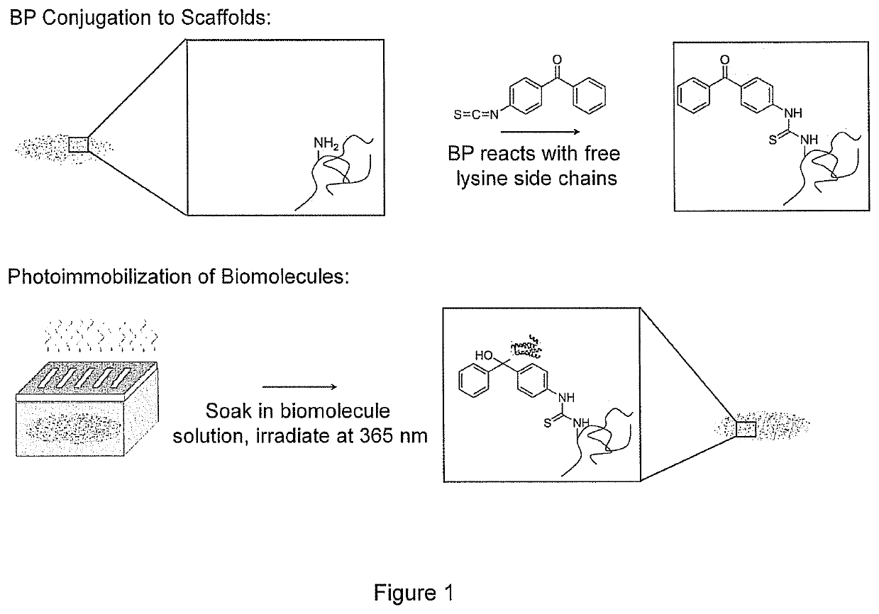
Attachment of BP to CG Scaffolds

[0069]A 20 mM solution of benzophenone-4-isothiocyanate containing 0.5 M N,N-diisopropylethylamine was prepared in dimethyl formamide (DMF) via an established method [36]. CG scaffolds were added to the solution and allowed to react at room temperature in the dark for 48 hrs. Scaffolds were thoroughly rinsed three times in DMF, 200 proof ethanol, and water (ELGA LabWater Reservoir, Veolia Water Systems, Buckinghamshire, UK) each for 1 hr; BP-tethered CG scaffolds were then stored under water and in the dark.

example 3

chment of Biomolecules onto BP-Presenting CG Scaffolds

[0070]Biotinylated concanavalin A (ConA-biotin) was purchased from Vector Laboratories (Burlingame, Calif.), fibronectin (FN) was purchased from Invitrogen (Carlsbad, Calif.) and N-Cadherin (NC) was purchased from R&D Systems (Minneapolis, Minn.). Stock solutions of biomolecules were prepared by resuspending the lyophilized proteins in the manufacturer's recommended buffer solutions to a concentration of 1 mg / mL. For ConA-biotin and FN, the buffer was phosphate buffered saline, pH=7.4 (PBS); PBS containing Ca2+ and Mg2+ was used for NC. The solutions were aliquotted and stored at −20° C. Immediately prior to use, the stock solutions were diluted in the respective buffers to yield the empirically determined final solution concentrations of 5 μg / mL ConA-biotin or 100 μg / mL FN.

[0071]BP-modified scaffolds were placed onto a microscope slide and surrounded by a rubber o-ring. A 20 μL aliquot of protein solution was added to the top of...


PUM

PropertyMeasurementUnit
at a wavelengthaaaaaaaaaa
a wavelengthaaaaaaaaaa
porosityaaaaaaaaaa

Abstract

The invention provides methods and compositions for making and using collagen-glycosaminoglycan three-dimensional scaffolds immobilized with biomolecules that are spatially and temporally patterned. The method comprises adding benzophenone to a collagen-glycosaminoglycan three dimensional scaffold in the dark; adding one or more biomolecules to one or more areas of the collagen-glycosaminoglycan three-dimensional scaffold (which can be done optionally in the dark or in the light); and exposing the collagen-2glycosaminoglycan three-dimensional scaffold to light at a wavelength of about 350 to about 365 nm.

Description

PRIORITY[0001]This application claims the benefit of U.S. Ser. No. 61 / 446,794, filed Feb. 25, 2011, which is incorporated herein by reference in its entirety.GOVERNMENT INTEREST[0002]This invention was made with government support under contract number NIH NIGMS T32GM070421, DE-FG02-07ER46453, and DE-FG02-07ER46471 awarded by the United States National Institutes of Health and the United States Department of Energy. The United States government has certain rights in the invention.BACKGROUND OF THE INVENTION[0003]The extracellular matrix (ECM) is a complex organization of structural proteins such as collagens and proteoglycans. A wide variety of tissue engineering scaffolds have been created in attempts to mimic features of the native ECM. Notably, microstructural features [1-3], mechanical properties [4-6], and inclusion of soluble or insoluble biomolecules [7, 8] have all been shown to significantly influence cell behaviors such as adhesion, growth, and differentiation as well as t...

Claims


Application Information

Patent Timeline
Patent Type & Authority Patents(United States)
IPC IPC(8): A61L27/38A61K35/12A61L27/50C12N5/077C12N5/00C08B37/00A61L27/56A61K35/32A61L27/54A61L27/26A61L27/48A61K38/18A61K38/30
CPCC08B37/0063A61L27/38C12N5/0658C12N5/0062A61L27/54C12N5/0657A61L27/56A61L27/26A61K35/12C12N5/0654A61K35/32A61L27/48A61L27/50C12N5/0655C08L89/06C12N2533/54A61L2430/06A61L2430/02A61L2430/30A61L2430/40A61L2430/20A61K38/30A61K38/1858A61L2430/32A61L2430/28
Inventor BAILEY, RYAN C.HARLEY, BRENDAN A.MARTIN, TERESA A.CALIARI, STEVEN R.
Owner THE BOARD OF TRUSTEES OF THE UNIV OF ILLINOIS
Why Eureka
Try Eureka