Methods for detecting protein-protein interactions

a protein-protein interaction and kit technology, applied in the field of protein-protein interaction detection kits, can solve the problems of lack of resolution for imaging subcellular structures, and the scheme confers a great potential for false positive interactions

Active Publication Date: 2020-09-22
INST NAT DE LA SANTE & DE LA RECHERCHE MEDICALE (INSERM) +1
View PDF4 Cites 0 Cited by
  • Summary
  • Abstract
  • Description
  • Claims
  • Application Information

AI Technical Summary

Benefits of technology

[0081]In some embodiments, the screening method of the present invention is particularly suitable for screening upstream modulators of the binding between the polypeptide (A) and the polypeptide (B). Actually, the candidate compound may indirectly modulate the binding between the 2 polypeptides (A) and (B) by interacting with an upstream component that will then modulate the binding. For example, focused kinase inhibitor compound libraries may be used to provide the candidate compounds. Thus, the candidate compound may be a compound already identified as a protein kinase inhibitor. These may be used to identify compound ‘hits’ (typically non-selective, low potency chemical start points). Available sequence knowledge, structural predictions and known kinase ligands may also be used to virtually screen commercially available compounds for novel compound hits. In some embodiments, the candidate compounds may inhibit or down-regulate the expression of a particular host cell gene, including coding and non coding sequence (e.g. miRNA). Such candidate compounds may comprise nucleic acid for example, oligonucleotide sequences, specifically designed to inhibit the expression of one or more host cell sequences—. Suitable candidate compounds may include, for example, DNA or RNA oligonucleotides, preferably antisense oligonucleotides. Such siRNA oligonucleotides may take the form of single or double-stranded RNA molecules which have been modified in some way (for example by chemical modification) to be nuclease resistant. In order to decrease or down-regulate the expression of a particular host cell sequence, or to block the activity of the host cell sequence (e.g. miRNA), the host cell may be contacted or transfected with any of the abovementioned candidate compounds. By analysing native or wild-type host cell sequences and with the aid of algorithms such as BIOPREDsi, one of skill in the art could easily determine or computationally predict nucleic acid sequences that have an optimal knockdown effect for these genes (see for example: http: / / www.biopredsi.org / start.html). Accordingly, the skilled man may generate and test an array or library of different oligonucleotides to determine whether or not they are capable of modulating the expression, function and / or activity of certain host cell sequence.
[0084]In some embodiments, the screening method of the present invention is performed in a high throughput screening assay. High-throughput screening (HTS) assays are typically used in drug discovery. Using robotics, data processing and control software, liquid handling devices, and sensitive detectors, High-throughput screening allows a quickly conduct millions of chemical, genetic, or pharmacological tests. Through this process one can rapidly identify the candidate compounds capable of modulating the binding between polypeptides (A) and (B).

Problems solved by technology

The method benefits from a great sensitivity due to the enzymatic activity of the luciferase reporter, but suffers from a lack of resolution for imaging subcellular structures.
However, this scheme confers a great potential of false positive interactions due to spontaneous co-folding of split-proteins moieties.

Method used


Image

  • Methods for detecting protein-protein interactions
  • Methods for detecting protein-protein interactions
  • Methods for detecting protein-protein interactions

Examples


example

[0090]Introduction:

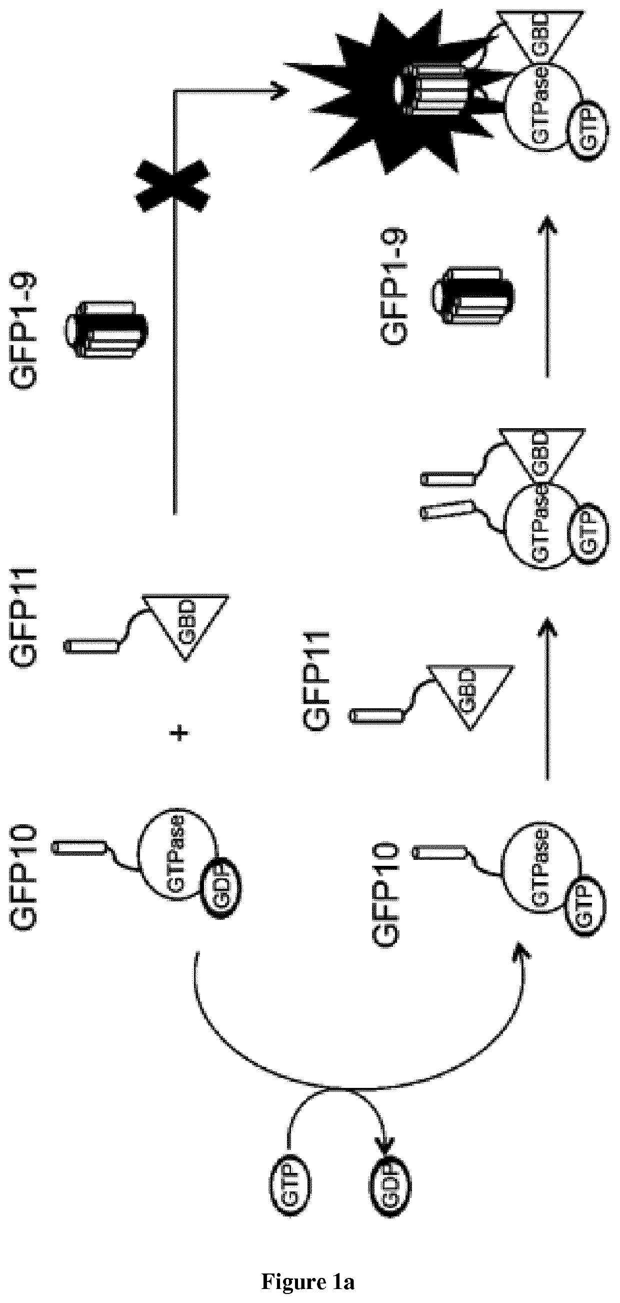
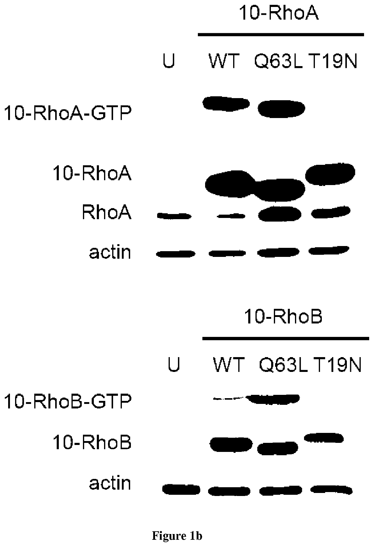
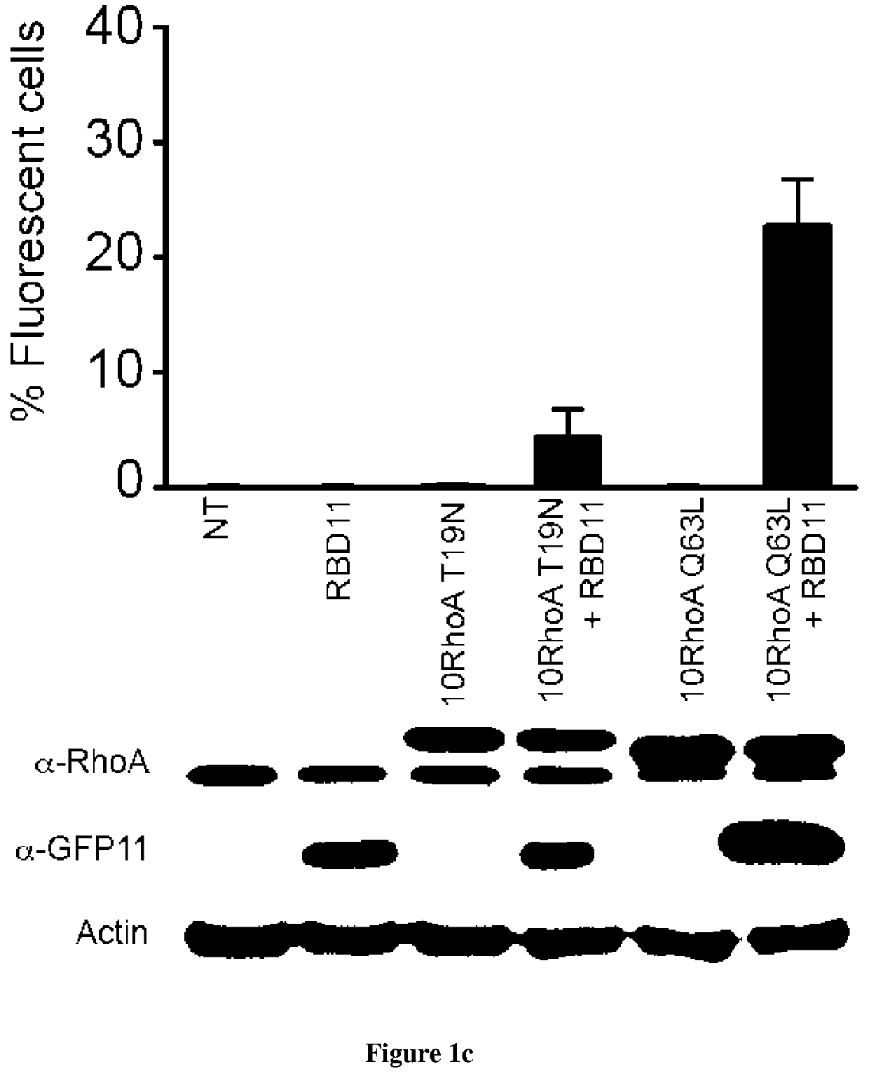
[0091]Small GTPases play an important role in signal transduction via transmembrane receptors to drive cytoplasmic or nuclear responses. They are involved in various fundamental cellular processes ranking from cytoskeleton organization to cell migration, and therefore are compelling pharmacological targets. The human Ras superfamily consists of 154 members divided in five main families: Ras, Rho, Rab, Arf and Ran. The Ras family is composed of three members H-Ras, K-Ras and N-Ras that are very closely related, with 85% amino acid sequence identity. Rho family proteins regroup small GTPases that contain a conserved Rho insert domain in the GTPase domain16. The leader members in the Rho subfamilies are RhoA, Rac1 and Cdc42 GTPases. Ras and Rho GTPases are molecular switches that cycle between GTP and GDP bound states. The activation state of Ras and Rho proteins depends on whether they are bound to GTP (active) or GDP (inactive). Binding to GTP is promoted by Rho Gu...


PUM

PropertyMeasurementUnit
concentrationsaaaaaaaaaa
fluorescenceaaaaaaaaaa
fluorescentaaaaaaaaaa

Abstract

The present invention relates to methods and kits for detection protein-protein interactions. In particular, the present invention relates to a method for detecting the binding between a first polypeptide (A) and a second polypeptide (B) in a cell comprising i) providing a cell that expresses (a) a polypeptide (GFP1-9) comprising an amino acid sequence having at least 90% of identity with the amino acid sequence selected from the group consisting of SEQ ID NO: 1-4 (b) a first fusion protein wherein the polypeptide (A) is fused to a polypeptide (GFP10) having an amino an amino acid sequence having at least 90% of identity with the amino acid sequence selected from the group consisting of SEQ ID NO:5-7 (c) a second fusion protein wherein the polypeptide (B) is fused to a polypeptide (GFP11) having an amino an amino acid sequence having at least 90% of identity with the amino acid sequence selected from the group consisting of SEQ ID NO: 8-9 and (d) an intrabody specific for the complex formed by the self-assembly of the first, second and third polypeptides (a), (b) and (c) ii) detecting the fluorescence wherein when the fluorescence is detected it is concluded that the polypeptide (A) binds to polypeptide (B) and wherein the fluorescence is not detected it is concluded that the polypeptides (A) does not bind to polypeptide (B).

Description

FIELD OF THE INVENTION[0001]The present invention relates to methods and kits for detection protein-protein interactions.BACKGROUND OF THE INVENTION[0002]Most cellular functions are driven by protein-protein interactions that regulate signal transduction and gene expression. Identifying partners involved in signaling pathways is of key importance in elucidating fundamentals of gene regulation mechanisms and in identifying aberrant cellular signaling processes occurring in various diseases. In recent years, a growing number of protein-based fluorescent biosensors have been developed to quantify and localize these interactions in living cells1. Fluorescence and Bioluminescence Resonance Energy transfer (FRET and BRET) based biosensors have emerged rapidly as they enable dynamic observations of protein-protein interactions2. Another class of reporters known as Protein-fragment Complementation Assay (PCA) has been developed to monitor direct interactions. These include split reporters f...

Claims


Application Information

Patent Timeline
Patent Type & Authority Patents(United States)
IPC IPC(8): G01N33/68G01N33/542G01N33/50G01N33/533C07K16/18C40B30/04
CPCG01N33/542C07K16/18G01N33/533C40B30/04G01N33/6845G01N33/5008C07K2319/70
Inventor CABANTOUS, STÉPHANIEKORAÏCHI, FATENFAVRE, GILLES
Owner INST NAT DE LA SANTE & DE LA RECHERCHE MEDICALE (INSERM)
Why Eureka
Try Eureka