Systems and methods to deliver point of care alerts for radiological findings

a technology of alerts and point of care, applied in the field of improved learning systems and methods for medical image processing, can solve the problems of less staff, less skilled staff, and difficulty in effective management and use of imaging and information systems for examination, diagnosis, and treatment of patients, and achieve the effect of improving imaging quality control

Active Publication Date: 2020-09-22
GENERAL ELECTRIC CO
View PDF59 Cites 148 Cited by
  • Summary
  • Abstract
  • Description
  • Claims
  • Application Information

AI Technical Summary

Problems solved by technology

A variety of economy, operational, technological, and administrative hurdles challenge healthcare facilities, such as hospitals, clinics, doctors' offices, imaging centers, teleradiology, etc., to provide quality care to patients.
Economic drivers, less skilled staff, fewer staff, complicated equipment, and emerging accreditation for controlling and standardizing radiation exposure dose usage across a healthcare enterprise create difficulties for effective management and use of imaging and information systems for examination, diagnosis, and treatment of patients.
Healthcare provider consolidations create geographically distributed hospital networks in which physical contact with systems is too costly.
Healthcare provider (e.g., x-ray technologist, doctor, nurse, etc.) tasks including image processing and analysis, quality assurance / quality control, etc., are time consuming and resource intensive tasks impractical, if not impossible, for humans to accomplish alone.

Method used


Image

  • Systems and methods to deliver point of care alerts for radiological findings
  • Systems and methods to deliver point of care alerts for radiological findings
  • Systems and methods to deliver point of care alerts for radiological findings

Examples


example imaging

Systems

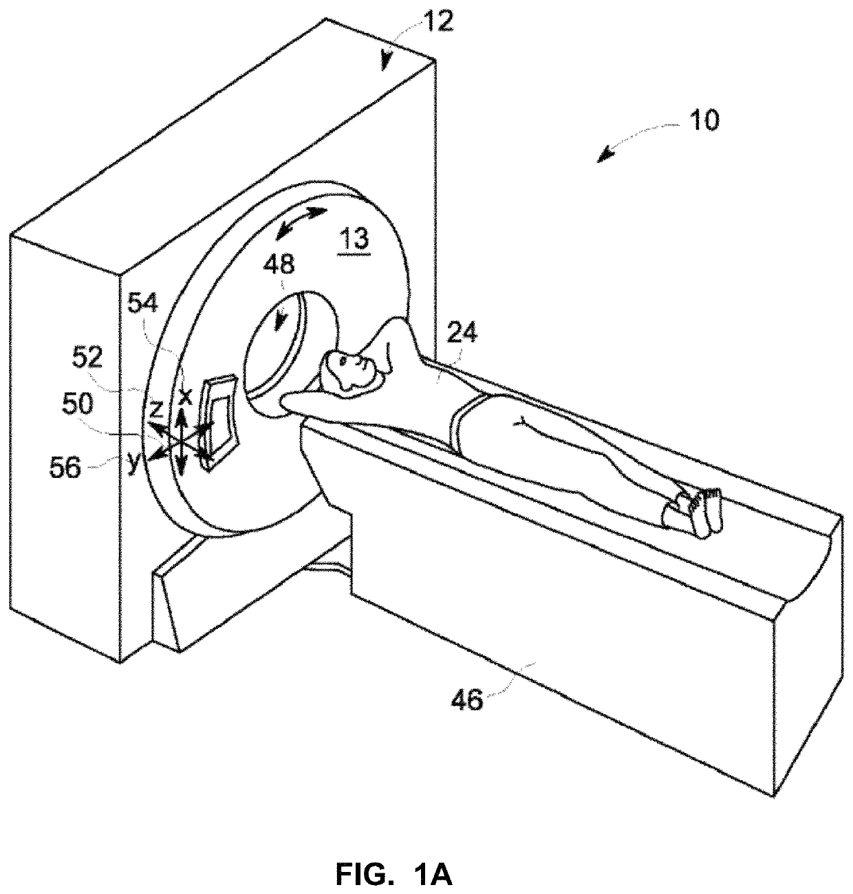
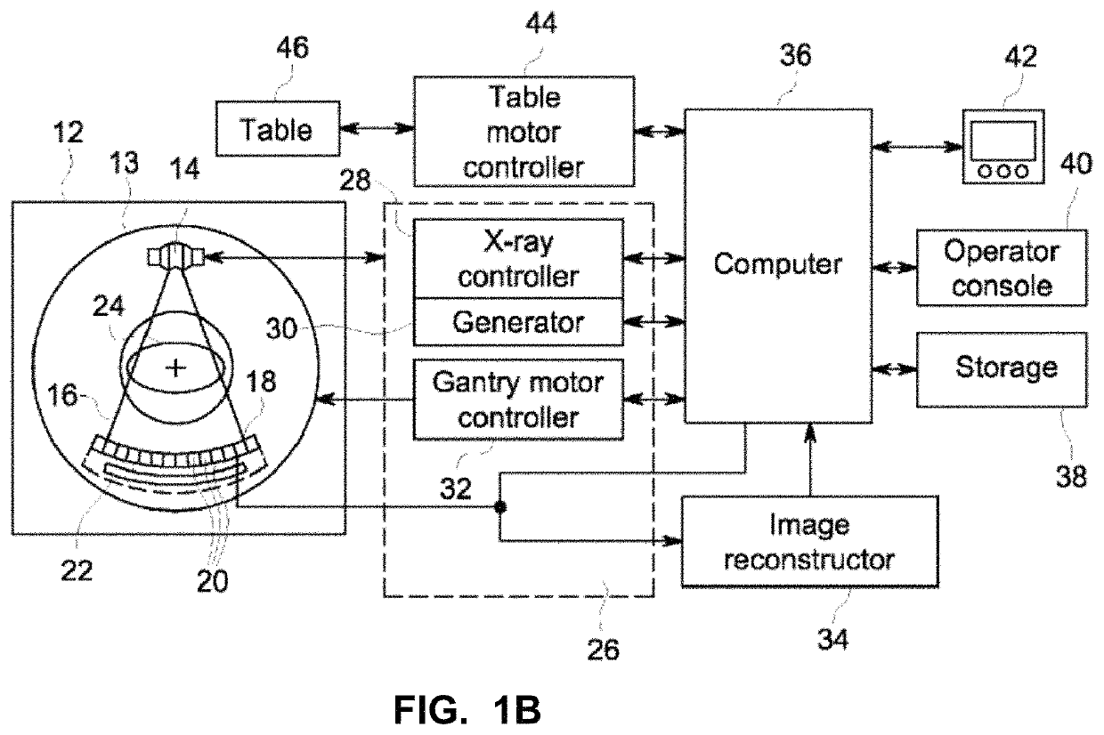
[0060]The methods, apparatus, and articles of manufacture described herein can be applied to a variety of healthcare and non-healthcare systems. In one particular example, the methods, apparatus, and articles of manufacture described herein can be applied to the components, configuration, and operation of a computed tomography (CT) imaging system. FIGS. 1A-1B illustrate an example implementation of a CT imaging scanner to which the methods, apparatus, and articles of manufacture disclosed herein can be applied. FIGS. 1A and 1B show a CT imaging system 10 including a gantry 12. Gantry 12 has a rotary member 13 with an x-ray source 14 that projects a beam of x-rays 16 toward a detector assembly 18 on the opposite side of the rotary member 13. A main bearing may be utilized to attach the rotary member 13 to the stationary structure of the gantry 12. X-ray source 14 includes either a stationary target or a rotating target. Detector assembly 18 is formed by a plurality of detector...

example learning

Network Systems

[0067]FIG. 3 is a representation of an example learning neural network 300. The example neural network 300 includes layers 320, 340, 360, and 380. The layers 320 and 340 are connected with neural connections330. The layers 340 and 360 are connected with neural connections 350. The layers 360 and 380 are connected with neural connections 370. Data flows forward via inputs 312, 314, 316 from the input layer 320 to the output layer 380 and to an output 390.

[0068]The layer 320 is an input layer that, in the example of FIG. 3, includes a plurality of nodes 322, 324, 326. The layers 340 and 360 are hidden layers and include, the example of FIG. 3, nodes 342, 344, 346, 348, 362, 364, 366, 368. The neural network 300 may include more or less hidden layers 340 and 360 than shown. The layer 380 is an output layer and includes, in the example of FIG. 3, a node 382 with an output 390. Each input 312-316 corresponds to a node 322-326 of the input layer 320, and each node 322-326 o...


PUM


Abstract

Apparatus, systems, and methods to improve imaging quality control, image processing, identification of findings, and generation of notification at or near a point of care are disclosed and described. An example imaging apparatus includes a processor to at least: evaluate first image data with respect to an image quality measure; when the first image data satisfies the image quality measure, process the first image data using a trained learning network to generate a first analysis of the first image data; identify a clinical finding in the first image data based on the first analysis; compare the first analysis to a second analysis, the second analysis generated from second image data obtained in a second image acquisition; and, when comparing identifies a change between the first analysis and the second analysis, trigger a notification at the imaging apparatus regarding the clinical finding to prompt a responsive action.

Description

CROSS-REFERENCE TO RELATED APPLICATIONS[0001]This patent arises from U.S. patent application Ser. No. 15 / 821,161, which was filed on Nov. 22, 2017. U.S. patent application Ser. No. 15 / 821,161 is hereby incorporated herein by reference in its entirety. Priority to U.S. patent application Ser. No. 15 / 821,161 is hereby claimed.FIELD OF THE DISCLOSURE[0002]This disclosure relates generally to improved medical systems and, more particularly, to improved learning systems and methods for medical image processing.BACKGROUND[0003]A variety of economy, operational, technological, and administrative hurdles challenge healthcare facilities, such as hospitals, clinics, doctors' offices, imaging centers, teleradiology, etc., to provide quality care to patients. Economic drivers, less skilled staff, fewer staff, complicated equipment, and emerging accreditation for controlling and standardizing radiation exposure dose usage across a healthcare enterprise create difficulties for effective managemen...

Claims


Application Information

Patent Timeline
Patent Type & Authority Patents(United States)
IPC IPC(8): G06T7/00G16H50/20G16H10/60G16H30/40G06T7/70G16H40/63
CPCG16H40/63G16H10/60G16H30/40G16H50/20G06T7/0014G06T7/70G06T2207/10104G06T2207/10088G06T2207/20081G06T2207/10132G06T2207/30004G06T2207/10081G06T2207/10116G06T2207/30168G06T2207/20084
Inventor NYE, KATELYN ROSERAO, GIREESHA
Owner GENERAL ELECTRIC CO
Why Eureka
Try Eureka