
Intranasal stimulation for treatment of meibomian gland disease and blepharitis

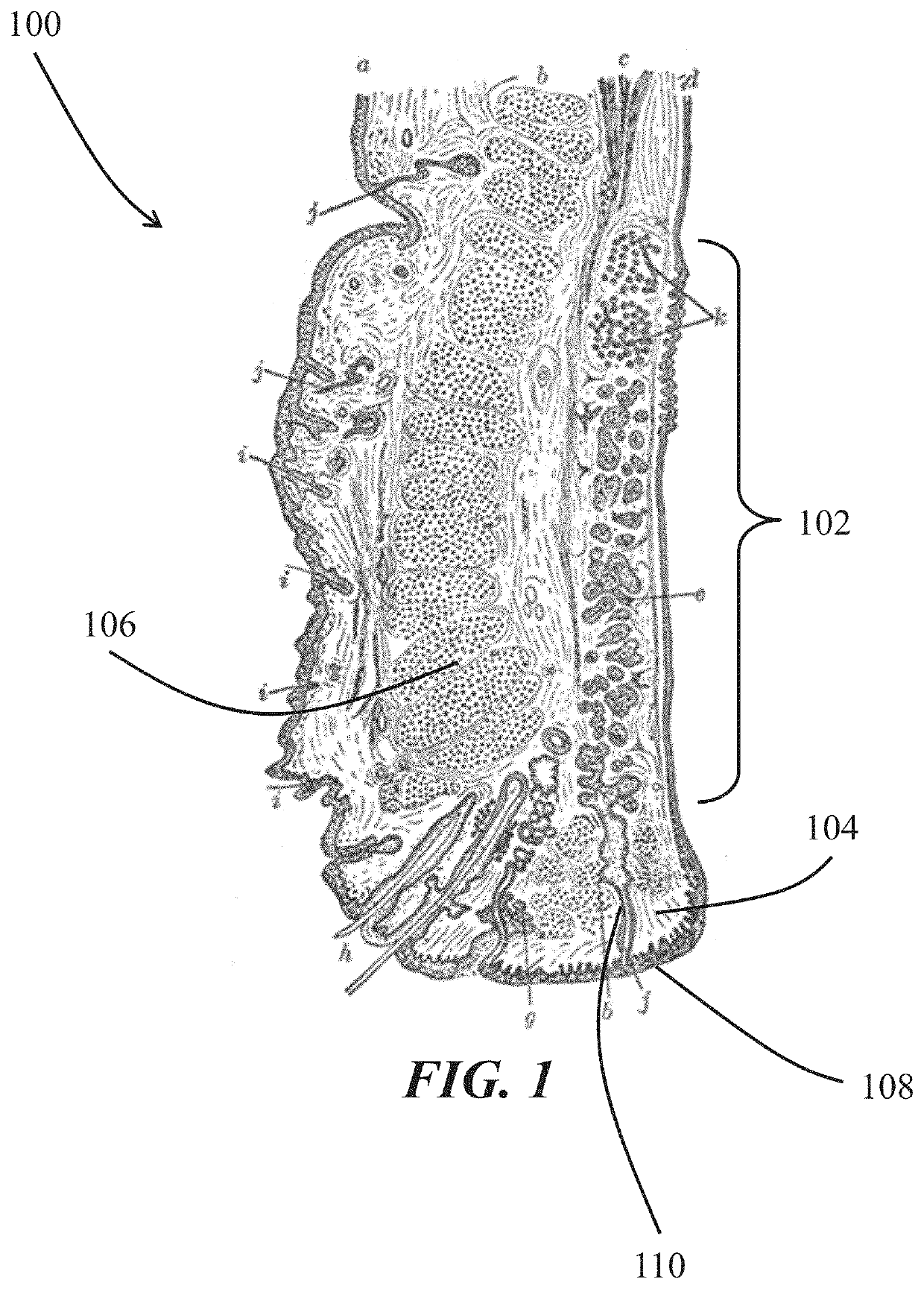
a meibomian gland and intranasal stimulation technology, applied in the field of intranasal stimulation for the treatment/or blepharitis, can solve the problems of increased risk of meibomian gland disease/dysfunction, increased tear evaporation, hyperocularity of tears, etc., to improve the secreted meibum quality and improve the effect of secreted meibum quality

Active Publication Date: 2021-02-16
OCULEVE
View PDF596 Cites 1 Cited by
  • Summary
  • Abstract
  • Description
  • Claims
  • Application Information

AI Technical Summary

Benefits of technology

[0006]In the methods described herein, the intranasal electrical stimulation may be delivered using a stimulator comprising at least one nasal insertion prong. In some variations the stimulator may comprise two nasal insertion prongs. A portion of a nasal insertion prong may be placed in contact with the nasal mucosa. In some variations, the portion of the nasal insertion prong placed in contact with the nasal mucosa may comprise a hydrogel electrode. The stimulator may in some variations have features to assist with hands-free or reduced-handling use. For example, the stimulator may comprise a strap to hold the nasal insertion prong in the nose. In some variations in which the stimulator comprises two nasal insertion prongs, the nasal insertion prongs may be biased toward each other. This bias may hold the stimulator in the nose during delivery of the intranasal electrical stimulation. In some variations, the intranasal electrical stimulation may be delivered using a stimulator that is temporarily attached to the nasal septum of the subject during delivery. In yet other variations, the intranasal electrical stimulation may be delivered by an implanted microstimulator. The implanted microstimulator may be implanted within a layer of submucosa.
[0010]Methods described herein may also treat a subject having meibomian gland disease and / or blepharitis by visualizing an eyelid margin of the subject during intranasal electrical stimulation of the subject, and adjusting at least one parameter of the intranasal electrical stimulation based on the visualization. The visualization may be performed with any suitable modality, such as a slit lamp or video capture. The intranasal electrical stimulation may increase secretion of meibum from the meibomian glands, such that the number of meibomian glands of the subject secreting meibum during the intranasal electrical stimulation is greater than the number of meibomian glands of the subject secreting meibum before the intranasal electrical stimulation. Adjusting at least one parameter of the intranasal electrical stimulation may comprise changing a first stimulation waveform to a second stimulation waveform. Changing from the first waveform to the second waveform may result in more uniform meibum expression between the two eyes of the subject. That is, the difference between the number of meibomian glands of the first eye of the subject secreting meibum and the number of meibomian glands of the second eye of the subject secreting meibum during intranasal electrical stimulation with the first stimulation waveform may be larger than the difference between the number of meibomian glands of the first eye secreting meibum and the number of meibomian glands of the second eye secreting meibum during intranasal electrical stimulation with the second stimulation waveform.

Problems solved by technology

As a result of these changes, a reduction in quantity and / or quality of meibum in the tear film layer may result in instability of the tear film, increased tear evaporation, hyperosmolarity of the tears, and / or alteration of the ocular surface.
The risk of meibomian gland disease / dysfunction may increase with certain risk factors, including age, reduced local androgen concentration, certain racial backgrounds, use of certain medications, wearing of contact lenses, and / or wearing of eye makeup.
Despite the prevalence of meibomian gland disease / dysfunction, however, these treatment approaches have had limited success.

Method used


Image

  • Intranasal stimulation for treatment of meibomian gland disease and blepharitis
  • Intranasal stimulation for treatment of meibomian gland disease and blepharitis
  • Intranasal stimulation for treatment of meibomian gland disease and blepharitis

Examples


example # 1

Example #1

Acute Meibum Secretion

[0096]A study was conducted to evaluate the effect of intranasal stimulation on meibum secretion in subjects with dry eye disease (DED) by visual assessment of various measures of meibomian gland function and assessment of tear meniscus height and tear meniscus area.

[0097]Twenty-five DED subjects were enrolled. Subjects were selected to have a baseline Ocular Surface Disease Index score of at least 13, with no more than three responses of “not applicable”; in at least one eye, a baseline Schirmer's test with anesthetic of less than or equal to 10 mm over 5 minutes, and in the same eye, a Schirmer's test that was at least 7 mm higher with cotton swab nasal stimulation; in at least one eye, a lower eyelid margin meibum quality global assessment score of greater than or equal to 1 and not non-expressible, where the score was based on the summed score for each of eight glands in the central third of the lower lid, where each gland was given a score of 0-3...

example # 2

Example #2

Meibomian Gland Morphology

[0105]A study was conducted explore the effect of intranasal stimulation on meibomian gland morphology. Anterior segment optical coherence tomography (AS-OCT) meibography was performed on the lower eyelids of each of twelve dry eye subjects before and after three minutes of intranasal stimulation. Intranasal stimulation was delivered using a handheld stimulator similar to the stimulator 400 shown in FIGS. 4A-4E and described in more detail with respect to Example #1. The area and perimeter of selected meibomian glands were quantified, and the effect of intranasal stimulation on meibomian glands was determined by morphological changes after stimulation. Within the selected images, three to four glands were analyzed. Central meibomian glands clearly shown in both sets of AS-OCT images were selected for analysis, according to the accuracy and quality of the image, and the same meibomian glands were selected before and after stimulation. The area and ...

example # 3

Example #3

Tear Composition

[0107]A study was conducted on subjects with dry eye disease to evaluate acute tear production and the relative concentration of total lipid in tears collected pre- and post- a single treatment period with intranasal stimulation. This study thus indirectly tested for increased meibum secretion after intranasal stimulation, since an increase in acute tear production with a sustained or increased concentration of lipids indicates increased absolute meibum secretion.

[0108]Subjects were selected to have a baseline Ocular Surface Disease Index score of at least 13, with no more than three responses of “not applicable”; and in at least one eye, a baseline Schirmer's test with anesthetic of less than or equal to 10 mm over 5 minutes, and in the same eye, a Schirmer's test that was at least 7 mm higher with cotton swab nasal stimulation. Fifty-five subjects were enrolled in the study, and forty-one subjects (26 females and 15 males; mean age of 61.6±15.9 years (26 ...


PUM


Abstract

Described here are devices, systems, and methods for treating meibomian gland disease and / or blepharitis by providing intranasal stimulation. Generally, the devices may deliver electrical stimulation to the nasal mucosa. Intranasal stimulation may unblock obstructed meibomian glands and increase the lipid content of tears, both acutely during stimulation and subsequent to intranasal stimulation. The methods may include an initial round of stimulation to unblock obstructed meibomian glands, and subsequent shorter rounds of stimulation to cause meibum secretion and maintain the open glands.

Description

CROSS-REFERENCE TO RELATED APPLICATIONS[0001]This application claims priority to U.S. Provisional Application No. 62 / 330,763, filed May 2, 2016, and titled “INTRANASAL STIMULATION FOR TREATMENT OF MEIBOMIAN GLAND DYSFUNCTION AND BLEPHARITIS,” and to U.S. Provisional Application No. 62 / 429,059, filed Dec. 1, 2016, and titled “INTRANASAL STIMULATION FOR TREATMENT OF MEIBOMIAN GLAND DYSFUNCTION AND BLEPHARITIS,” each of which is hereby incorporated by reference in its entirety.FIELD[0002]Described here are devices, systems, and methods for treating meibomian gland disease / dysfunction and / or blepharitis using intranasal stimulation.BACKGROUND[0003]The meibomian glands are large sebaceous glands located in the tarsal plates of the upper and lower eyelids. Typically, the glands generate lipids (meibum), which are excreted onto the eyelid margin through excretory ducts during muscular contractions from eyelid movement. In patients with meibomian gland disease / dysfunction, abnormalities of ...

Claims


Application Information

Patent Timeline
Patent Type & Authority Patents(United States)
IPC IPC(8): A61N1/00A61N1/36A61N1/05
CPCA61N1/36046A61N1/0546A61N1/3603A61N1/36014A61N1/36034
Inventor FRANKE, MANFREDSENCHYNA, DIANE MICHELLEHAMILTON, DANIEL N.
Owner OCULEVE
Learn More