

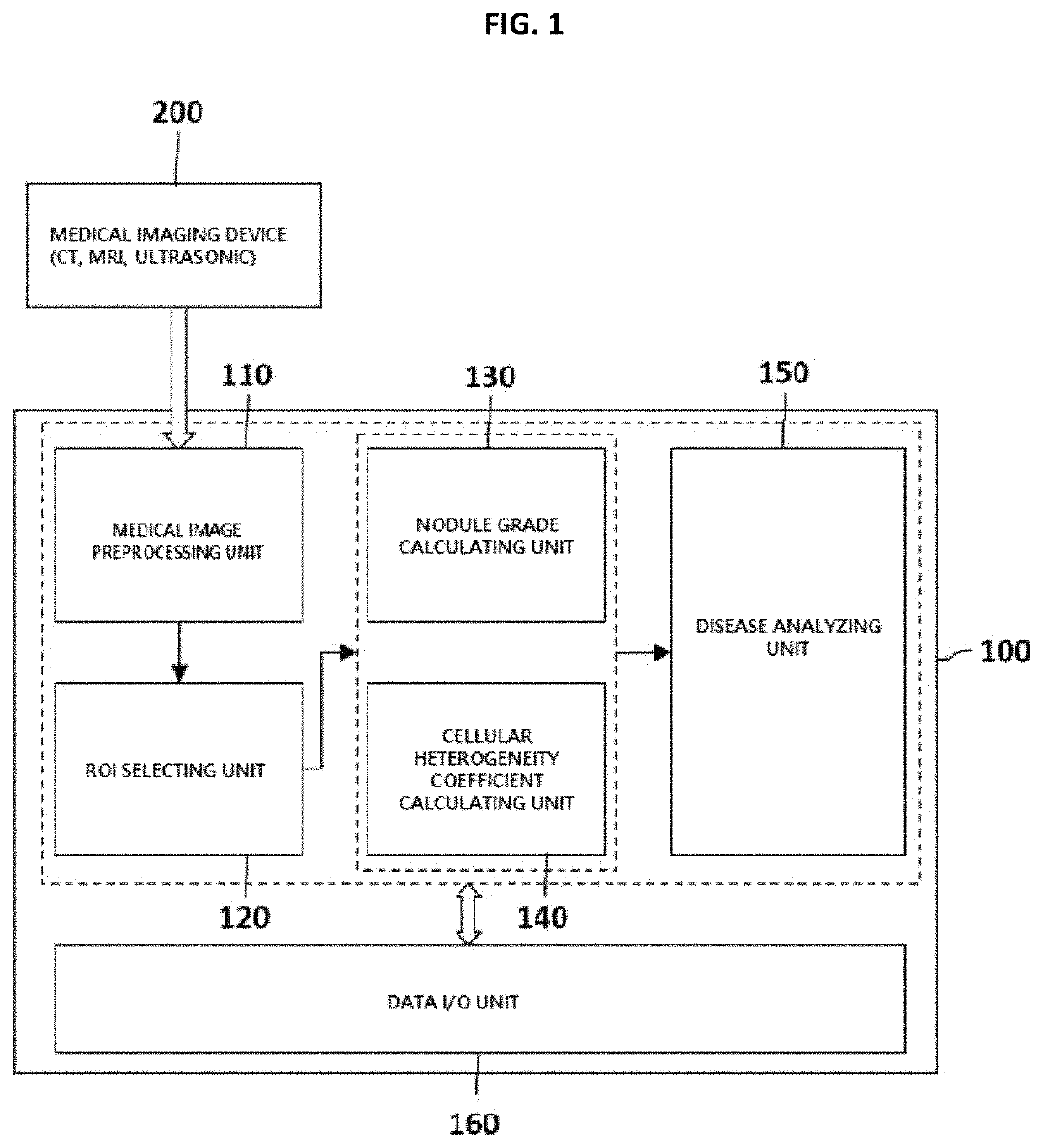
Method and apparatus for calculating abdominal disease diagnosis information based on medical image
a medical image and diagnosis information technology, applied in the field of medical image analysis methods and apparatus, can solve the problems of unnecessarily increasing the cost and time required for diagnosing the degree of disease of the patient, and liver cancer, and achieve the effect of reducing the cost and time required for diagnosis
- Summary
- Abstract
- Description
- Claims
- Application Information
AI Technical Summary
Benefits of technology
Problems solved by technology
Method used
Image

Examples
Embodiment Construction
[0025]The objects and effects of the present disclosure and the technical features for achieving them will become apparent with reference to the embodiments described in detail below along with the accompanying drawings. In the following description of the present disclosure, known functions or configurations will not be described in detail when it is determined that the gist of the present disclosure may be unnecessarily obscured thereby.
[0026]In addition, the following terms are defined in consideration of the functions in the present disclosure and may vary depending on the intention of a user or an operator, or the customs.
[0027]However, the present disclosure is not limited to the embodiments disclosed below, but may be implemented in various other ways. The embodiments are provided just for perfect explanation of the present disclosure and for allowing those of skilled in the art to completely understand the present disclosure, and the present disclosure is defined only by the...
PUM
 Abstract
Description
Claims
Application Information