System and method for a tissue resection margin measurement device

a tissue resection and margin measurement technology, applied in the field of surgery, can solve the problems of insufficient resection, poor long-term benefits, and difficulty in localizing the lesion and precise removal of the lesion, and achieve the effect of minimal invasiveness

Active Publication Date: 2022-02-01
THE BRIGHAM & WOMEN S HOSPITAL INC +1
View PDF6 Cites 0 Cited by
  • Summary
  • Abstract
  • Description
  • Claims
  • Application Information

AI Technical Summary

Benefits of technology

[0033]In one form of the invention, the system may be used to identify the location of a particular airway. In this form of the invention, the system comprises means for bronchoscopic positioning of a sensor into an airway of the lung. This bronchoscopic positioning of the sensor in an airway of the lung (e.g., by positioning the sensor on a bronchoscope or on a catheter within the brochoscope and advancing the bronchoscope into the airway of interest) can be used to define the lobar, segmental or subsegmental bronchus for surgery such as segmentectomy, lobectomy or wedge resection during the actual operation. This function can be independent of the lesion margin measurement, and the position of the sensor identifying the bronchus can be correlated with the position of another device (e.g., a surgical instrument) carrying another sensor so that the surgeon can define the correct bronchus for surgery from the chest side of the operation. Thus, in this form of the invention, one sensor is positioned on a bronchoscope or on a catheter placed within the bronchoscope which is inserted into a specific airway so as to define the location of that specific airway, and another sensor is positioned on a surgical instrument which is advanced for surgery from the chest side of the operation, with the system continuously tracking the position of the sensor on the surgical instrument vis-à-vis the position of the sensor on the bronchoscope, so that the surgeon can continuously track the location of the surgical instrument relative to the airway of interest (identified by the sensor on the bronchoscope), e.g., to target the airway identified by the sensor on the bronchoscope, to avoid the airway identified by the sensor on the bronchoscope, etc.

Problems solved by technology

Significant deformation of the tissue due to high viscoelasticity, physiological motion (such as collapsing of the lung, breathing or beating motion), or tissue manipulation can lead to difficulty in localizing the lesion and precise removal of the lesion.
As a result, this can lead to insufficient resection, lesion recurrence locally, or by metastasis (in cancer), and poorer long-term benefits compared with cases where a sufficient margin is obtained.
The orthopedic trauma alone presents considerable pain and it can complicate the recovery process with patients.
Thoracic pain of this magnitude also complicates the task of recovering a patient from general anesthesia since the body acclimates to forced ventilation and the pain can interrupt natural chest rhythm.
This is due, at least in part, to the fact that there are only a limited number of instruments designed specifically to facilitate thoracic procedures in this way.
In the conventional method of performing VATS, however, the lung is collapsed during surgery, leading to difficulty in precisely locating the lesion and determining the resection margins.
Additionally, palpation of lung tissue is not always possible (particularly in the case of smaller or early stage cancers) due to the minimally invasive approach to surgery.
Imprecise surgical resection could lead to incomplete resection and subsequent lesion recurrence.
However, identifying and resecting the entire lesion is a challenging task due to the highly deformable nature of the breast.

Method used


Image

  • System and method for a tissue resection margin measurement device
  • System and method for a tissue resection margin measurement device
  • System and method for a tissue resection margin measurement device

Examples


Embodiment Construction

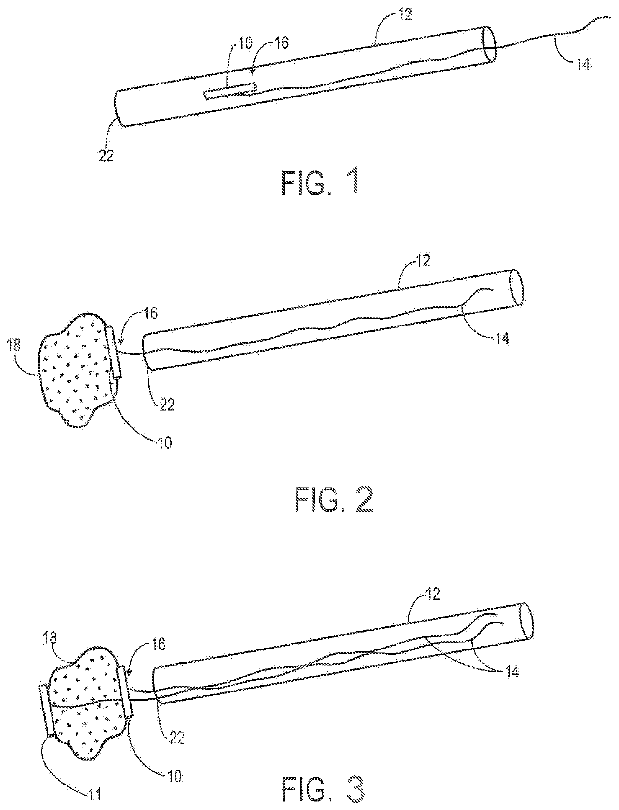
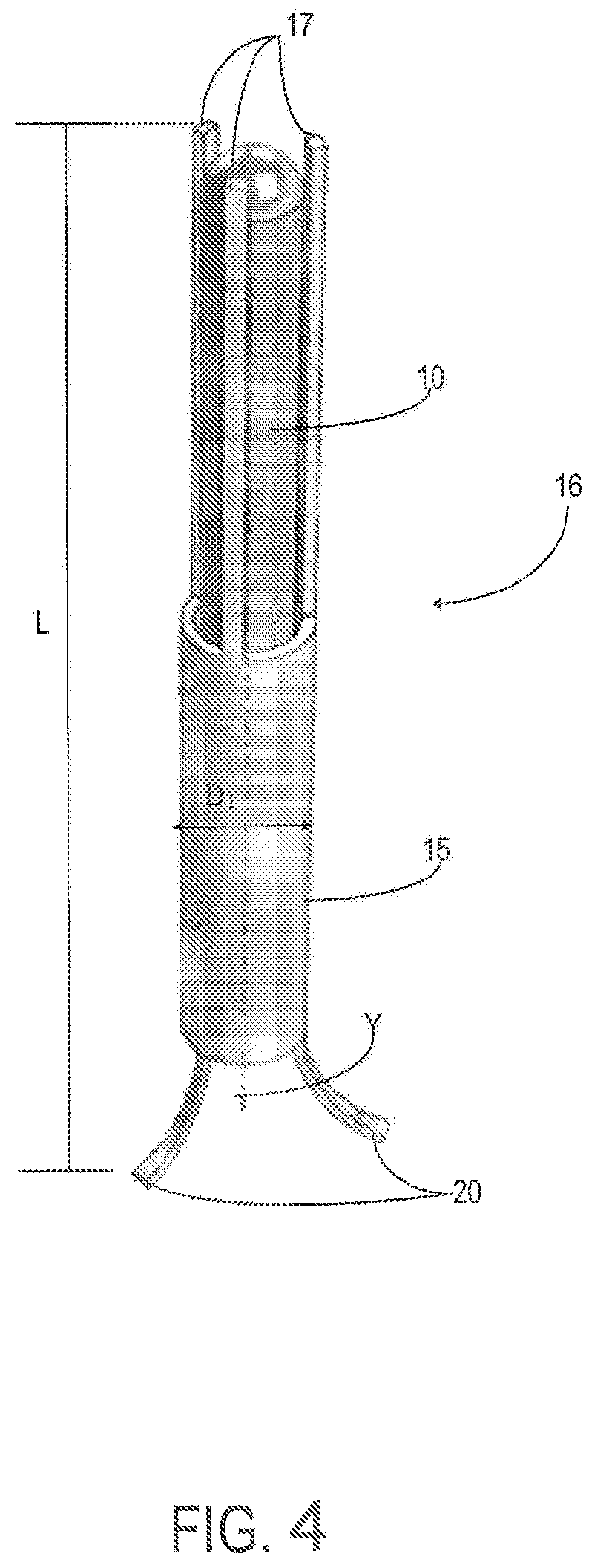
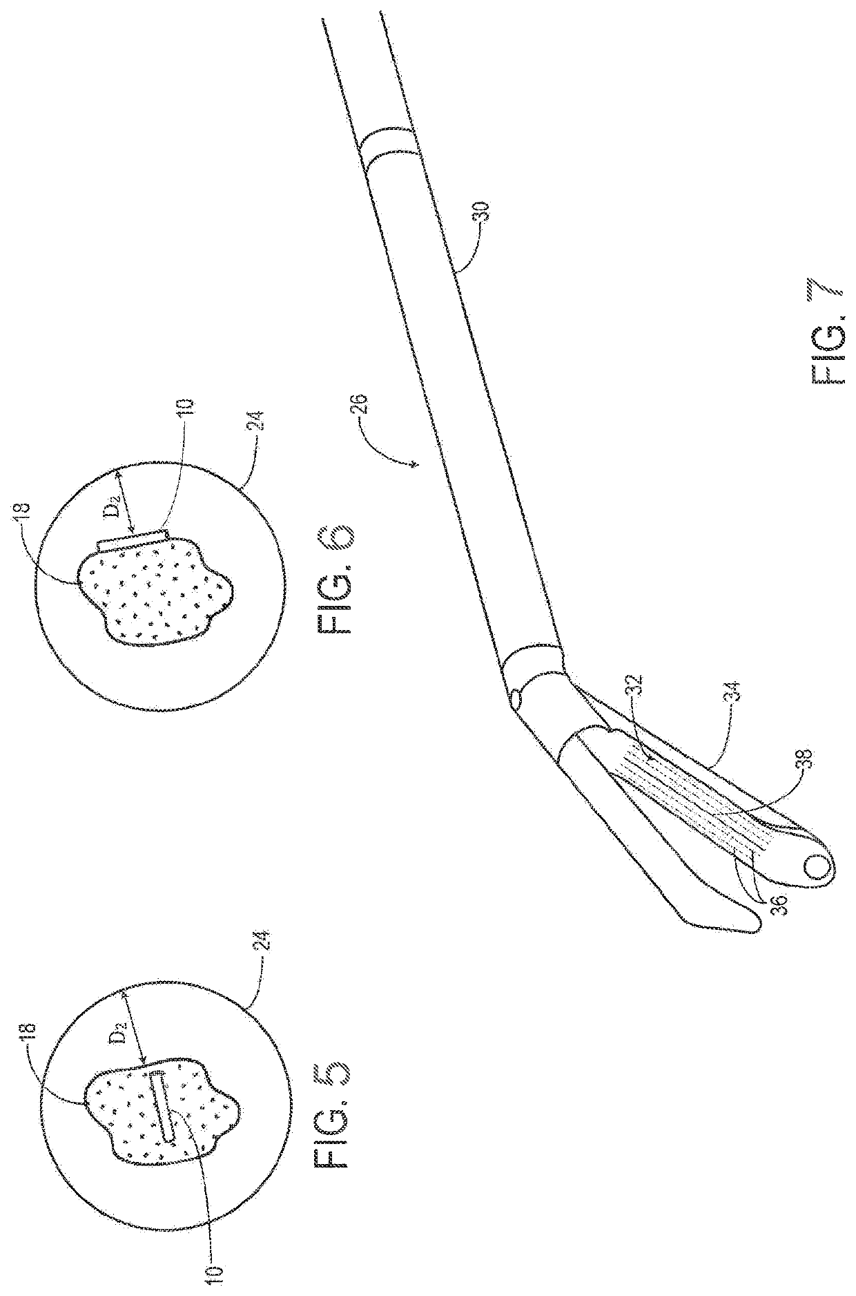
[0169]Before any embodiments of the invention are explained in detail, it is to be understood that the invention is not limited in its application to the specific details of construction and the arrangement of components set forth in the following description or illustrated in the following drawings. The invention is capable of other embodiments and of being practiced or of being carried out in other ways. Also, it is to be understood that the phraseology and terminology used herein is for the purpose of description and should not be regarded as limiting. The use herein of “including,”“comprising,” or “having” and variations thereof is meant to encompass the items listed thereafter and equivalents thereof as well as additional items. Unless specified or limited otherwise, the terms “mounted,”“connected,”“supported,” and “coupled” and variations thereof are used broadly and encompass both direct and indirect mountings, connections, supports, and couplings. Further, “connected” and “c...


PUM


Abstract

Embodiments of the invention provide a system and method for resecting a tissue mass. The system for resecting a tissue mass includes a first sensor for measuring a signal corresponding to the position and orientation of the tissue mass. The first sensor is dimensioned to fit inside of or next to the tissue mass. The system also includes a second sensor attached to a surgical instrument configured to measure the position and orientation of the surgical instrument. A controller is in communication with the first sensor and the second sensor, and the controller executes a stored program to calculate a distance between the first sensor and the second sensor. Accordingly, visual, auditory, haptic or other feedback is provided to the clinician to guide the surgical instrument to the surgical margin.

Description

REFERENCE TO PENDING PRIOR PATENT APPLICATIONS[0001]This patent application claims benefit of:[0002](1) prior U.S. Provisional Patent Application Ser. No. 62 / 923,137, filed Oct. 18, 2019 by Raphael Bueno et al. for SYSTEM AND METHOD FOR A TISSUE RESECTION MARGIN MEASUREMENT DEVICE; and[0003](2) prior U.S. Provisional Patent Application Ser. No. 63 / 054,921, filed Jul. 22, 2020 by Raphael Bueno et al. for SYSTEM AND METHOD FOR A TISSUE RESECTION MARGIN MEASUREMENT DEVICE.[0004]The two (2) above-identified patent applications are hereby incorporated herein by reference.FIELD OF THE INVENTION[0005]This invention relates to surgery in general, and more particularly to computer-assisted surgery.BACKGROUND OF THE INVENTION[0006]Minimally invasive surgical resection of lesions involves the precise excision of the lesion while sparing surrounding healthy and critical tissue. Some examples include, but are not limited to, breast conserving surgery and Video-Assisted Thoracic Surgery (VATS). S...

Claims


Application Information

Patent Timeline
Patent Type & Authority Patents(United States)
IPC IPC(8): A61B17/34A61B17/32A61B34/20A61B34/10A61B17/068A61B90/00A61B17/00
CPCA61B17/3468A61B17/068A61B17/320016A61B34/10A61B34/20A61B90/06A61B90/39A61B2017/008A61B2017/00119A61B2017/00809A61B2034/105A61B2034/2051A61B2090/067A61B2090/397A61B2090/3908A61B17/07207A61B2090/3912A61B2034/107A61B2017/07271A61B2017/00115A61B2017/2927A61B2090/3987A61B2090/3991
Inventor BUENO, RAPHAELJAGADEESAN, JAYENDERLUCAS, ALAN D.
Owner THE BRIGHAM & WOMEN S HOSPITAL INC
Why Eureka
Try Eureka