Composition and method for regulating the adhesion of cells and biomolecules to hydrophobic surfaces
a technology of hydrophobic surfaces and composition methods, applied in the field of biomolecules, can solve the problems of cell contact requirements, cell loss of natural architecture, and inability to function normally, and achieve the effect of not precisely mimicking the biological functions of cells
- Summary
- Abstract
- Description
- Claims
- Application Information
AI Technical Summary
Problems solved by technology
Method used
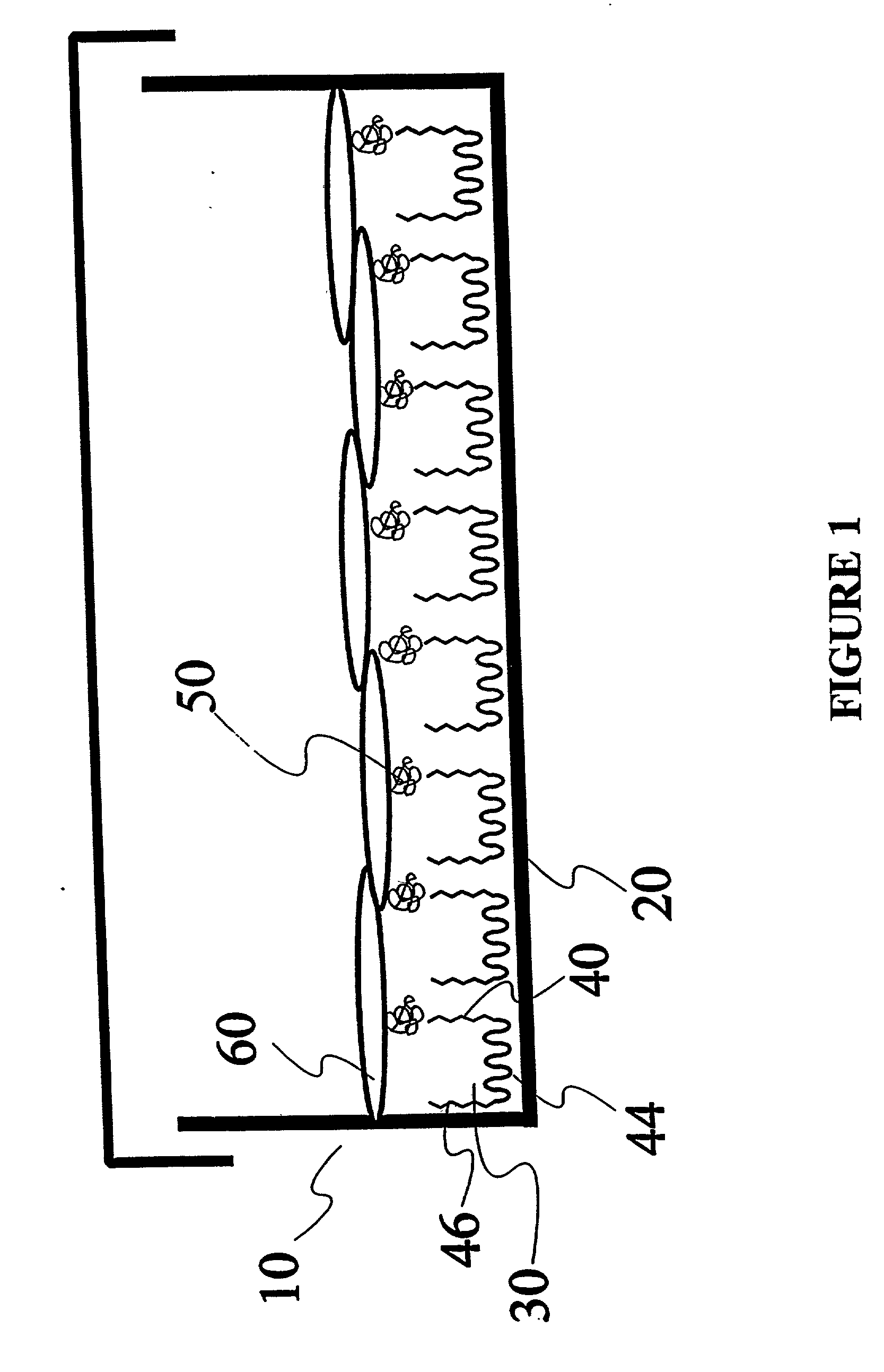
Image

Examples
example 1
Coupling of Amines to EGAP
[0123] (A) 1,3 Diaminopropane. 1,3 Diaminopropane (3.3 g) was mixed with 5 mL of deionized water. After the pH was adjusted to 8.2 with concentrated HCL, the solution was mixed with a solution of 0.5 g of 4-nitrophenyl chloroformate activated Pluronic.TM. F108 in 5.0 mL of deionized water. The reaction mixture, which immediately turned yellow, was kept at 25 C for 15 h. This solution was transferred to a dialysis tubing (with a molecular weight cutoff of 3500) and was dialyzed against 4 L of deionized water. During the 48 h dialysis process, water was changed five times until the low molecular weight material was assumed to be completely removed. The product was then recovered by lyophilization. The degree of substitution was determined by elemental nitrogen analysis. In this calculation the nitrogen content determined per a given mass of product was taken to exclusively derive from the attached diamine.
[0124] (B) 2-Aminoethanesulfonic Acid (Taurine). Tauri...
example 2
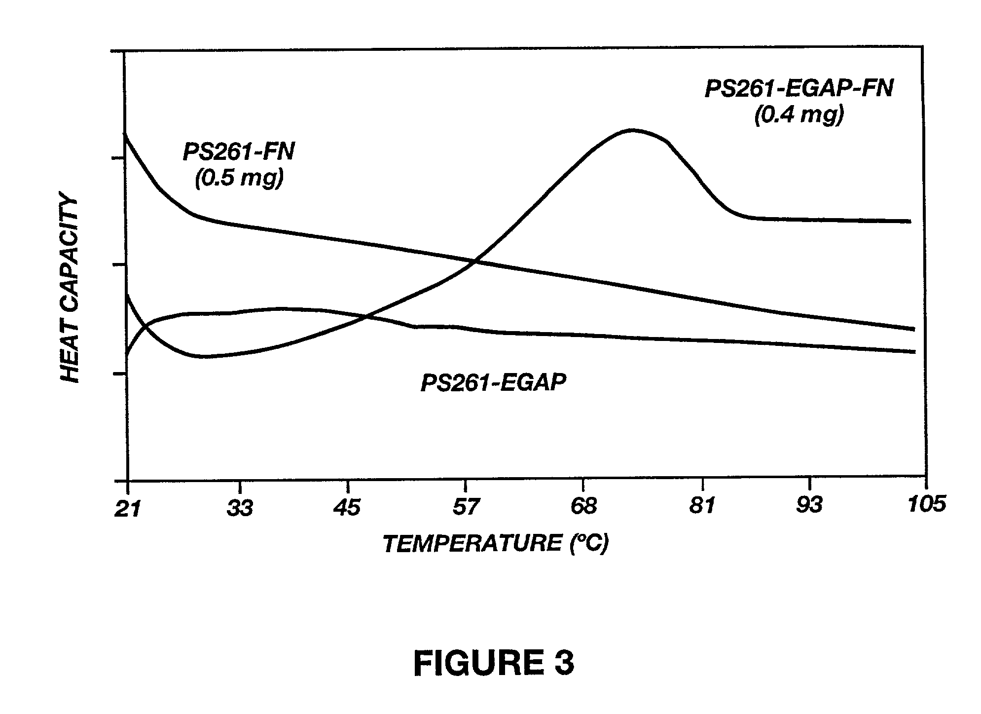
Calorimetric Observations of Fibronectin Conjugated to EGAP
[0126] PS latex particles with a diameter of 261 nm were purchased as a 10% (w / v) suspension were purchased from Seradyn, [State]. The block copolymer surfactant surfactant used was F108 having a molecular weight of 14600 were donated by BASF COW'S., [state]. Nsuccinimidyl-3-(2-pyridyl-dithiol) propionate (SPDP) was obtained from Pierce, [state]. Diihiothreitol (DTT) was from Bio-Rad, [state]. Fibronectin solution (FN, 1.5 mg / ml) was isolated from human plasma, and disposable prepacked PD-10 columns were purchased from Pharmacia, Wis.
[0127] The Pluronic.TM. F108-2-pyridyl disulfide derivative (GAP) was synthesized as described above.
[0128] Adsorption reaction was carried out in a mixture consisting of 10 .mu.L of the suspension of PS latex particles and 200 .mu.L of 0.5% (w / w) EGAP dissolved in deionized water. This mixture was incubated for 2 hours with shaking at room PS microspheres were washed and recovered by table cent...
example 3
Calorimetric Observations of Human Serum Albumin Conjugated to EGAP
[0132] Human Serum Albumin (HSA) conjugated EGAP was thiolated, purified, and conjugated to EGAP essentially as described in Example 2. The attachment of HSA to a PEO tether, already in place at the surface, was an obvious route to retention of structure, as seen in FIG. 4. The three traces in the FIG. 4 represent the thermograms for protein in solution, protein adsorbed onto bare PS particles, and proteins attached to the surface through the Pluronic.TM. F108 intermediate. As with fibronectin, all cooperative transitions are absent from the protein-particle adsorption complex. However, the PEO-tethered sample shows the characteristic complex melting curve of native HSA, reflecting the differential collapse of the three lobes of this protein. The transition enthalpies associated with thermal unfolding of HSA in the three different states are listed in Table 2. Due to the irreversible nature of these thermal transitio...
PUM
| Property | Measurement | Unit |
|---|---|---|
| pH | aaaaa | aaaaa |
| diameter | aaaaa | aaaaa |
| hydrophobic | aaaaa | aaaaa |
Abstract
Description
Claims
Application Information
 - R&D Engineer
- R&D Manager
- IP Professional
- Industry Leading Data Capabilities
- Powerful AI technology
- Patent DNA Extraction
Browse by: Latest US Patents, China's latest patents, Technical Efficacy Thesaurus, Application Domain, Technology Topic.
© 2024 PatSnap. All rights reserved.Legal|Privacy policy|Modern Slavery Act Transparency Statement|Sitemap