

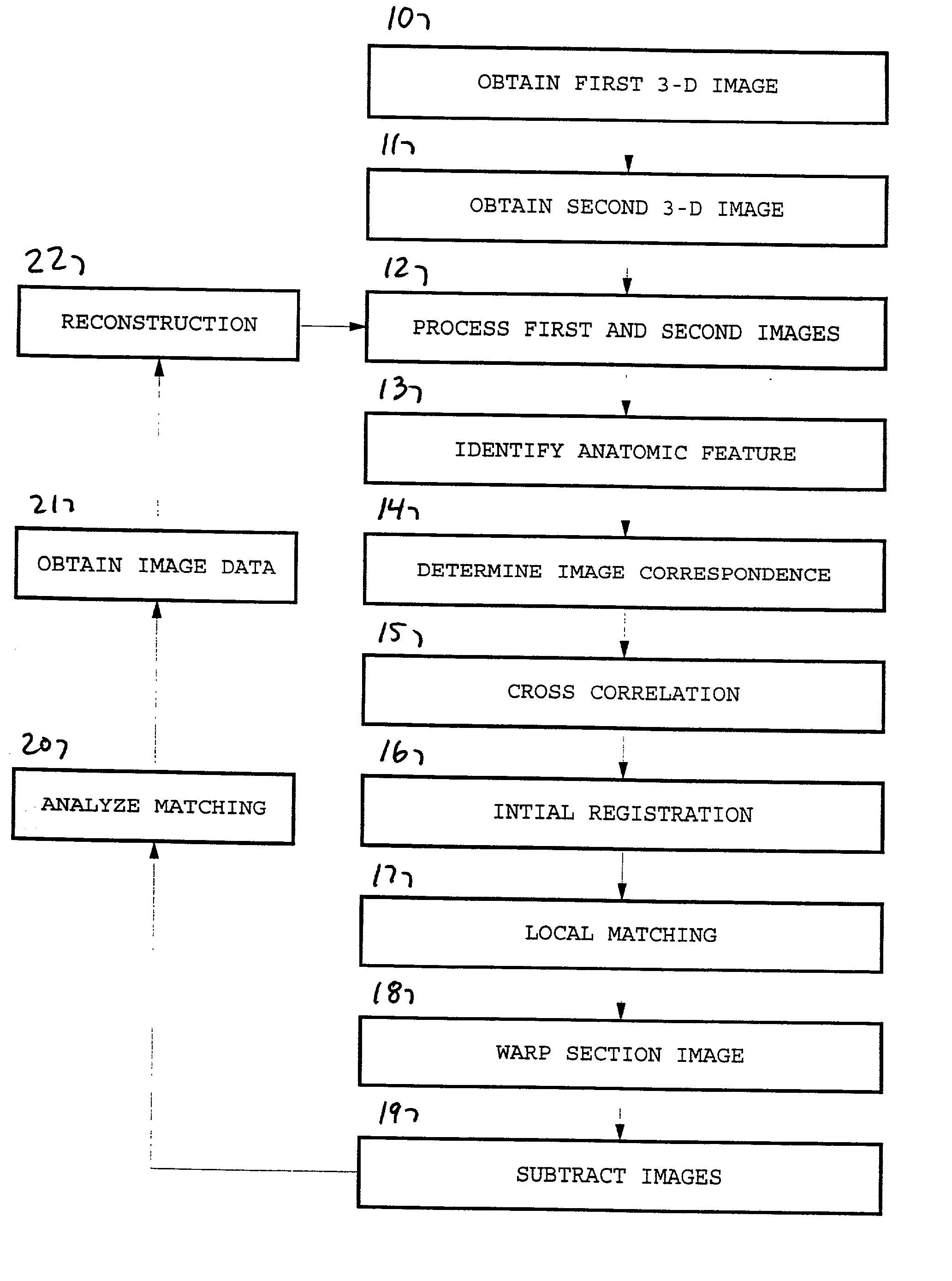
Method and system for the automated temporal subtraction of medical images
a technology of medical images and temporal subtraction, applied in the field of temporal analysis of medical images, can solve the problems of difficult and time-consuming for radiologists to compare current and previous, difficult to keep these positions unchanged in a clinical environment, and difficulty in viewing several images
- Summary
- Abstract
- Description
- Claims
- Application Information
AI Technical Summary
Benefits of technology
Problems solved by technology
Method used
Image

Examples
Embodiment Construction
[0071] A total of 14 cases (14 pairs of temporally sequential thoracic CT scans) were used to demonstrated the method according to the invention. The time interval between the current and previous thoracic CT scans was varied from two to seven months. The truth of the presence or absence of interval changes was obtained by an experienced radiologist reading the images. Two cases were reported to have interval changes, and the other cases had no interval changes, according to the radiologists.
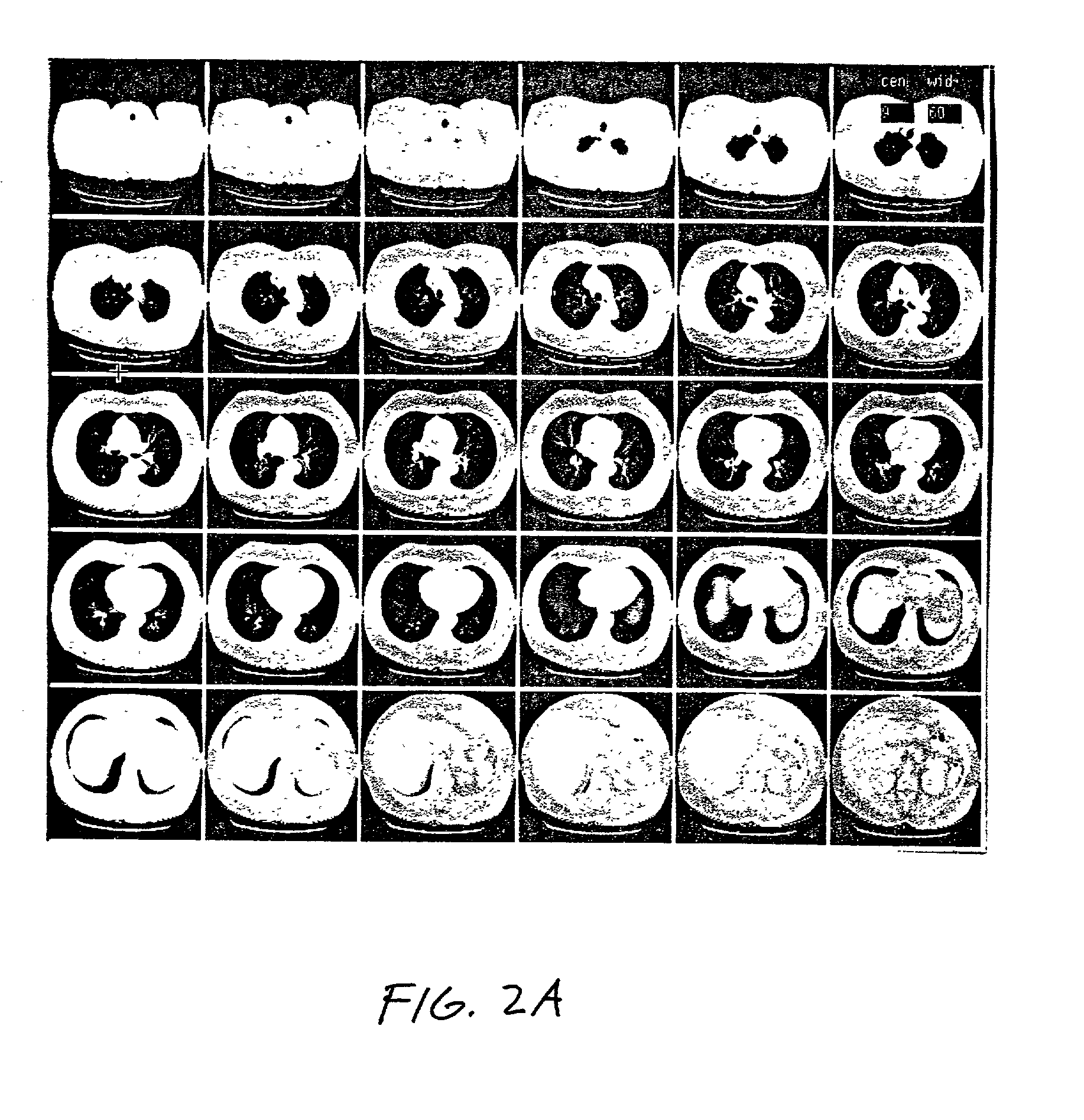
[0072] FIGS. 11A-11C show the case in which an anterior mediastinal mass (at the left side in the section images) increased in size from 3 cm in the previous scan to 4 cm in the current scan. On the subtracted section images (FIG. 11C), the dark area surrounding the mass indicates that the mass is increased in size. The dark area is indicated by the arrows. With the method according to the invention, the new findings or interval changes in the current sections are shown as dark patterns on the s...
PUM
 Abstract
Description
Claims
Application Information