Container uncapping mechanism and method

a container and uncapping technology, applied in mixer accessories, flat carrier supports, ultrafiltration, etc., can solve the problems of insufficient consistency, reliability, speed and automation of automated equipment for processing liquid-based specimens, and add time, material and labor costs to the processing required, etc., to meet current and projected needs in cancer screening and other cytology-based medical, analytical, screening and diagnostic procedures. , the effect of simple and inexpensive releasable coupling and avoiding contamination

Inactive Publication Date: 2003-05-15
HOLOGIC INC
View PDF9 Cites 11 Cited by
  • Summary
  • Abstract
  • Description
  • Claims
  • Application Information

AI Technical Summary

Benefits of technology

[0009] The processing assembly is coupled to a simple cover for the vial by means of a simple and inexpensive releasable coupling. When the cover is removed at the point-of-care site (physician's office, clinic, hospital, etc.), the processing assembly remains with the cover to allow medical personnel easy access to the container interior for insertion of a biological specimen into the vial. The cover, along with the attached processing assembly, is then replaced to seal the vial. The vial may then be sent to a laboratory for processing.
[0010] When the vial is manipulated in a simple way while still closed, the processing assembly detaches from the cover and remains in the vial for access by automated or manual laboratory equipment when the cover is subsequently removed. In a preferred embodiment, a downward force on the center of the cover is all that is required to detach the processing assembly from the cover. In contrast with the prior art specimen vials discussed above, the vial of the present invention requires no further interaction with the cover, which can be removed by a simple uncapping device and is discarded to avoid contamination. Ribs inside the vial support the processing assembly in the proper position for access during processing. This self-contained vial and processing assembly arrangement minimizes human operator exposure to biohazards, such as tuberculosis or other pathogens in sputum or in other specimens types, such as urine, spinal tap fluid, gastric washings, fine-needle aspirates, and gynecological samples.
[0013] Identifying molecular changes at the cellular level is one of the ways cancer can be detected early and at a more curable stage. Such molecular diagnostic devices can be used for early detection and diagnosis with the necessary sensitivity and specificity to justify their use as population-based screens for individuals who are at-risk for developing cancer. Such a molecular diagnostic device also can be used to characterize the tumor, thereby permitting the oncologist to stratify his / her patients, to customize therapy, and to monitor patients in order to assess therapeutic efficacy and disease regression, progression or recurrence. The availability of such tests will also foster the development of new and more effective therapeutic approaches for the treatment of early stage disease.
[0020] The filter assembly preferably has a holder, a frit seated in the holder, and a membrane filter positioned over and in contact with the outer surface of the frit. The frit can extend beyond the end of the holder. The membrane filter can be attached to the holder. The sidewall portion extending beyond the holder forms an area through which the specimen liquid can flow, creating a secondary flow. The holder can be configured so that the frit is slightly bowed outwardly at the center so that when pressure is applied to a slide during the specimen transferring step, the central portion of the frit flattens to more evenly contact the membrane filter to the slide for more effective transfer.

Problems solved by technology

In all of these endeavors, a limiting factor in the sample preparation protocol is adequately separating solid matter from its fluid carrier, and in easily and efficiently collecting and concentrating the solid matter in a form readily accessible to examination under a microscope.
All of this complexity adds time, and material and labor cost to the processing required prior to the actual cytology examination.
In general, automated equipment thus far developed for processing liquid-based specimens have not performed with sufficient consistency, reliability, speed and automation to satisfy current and projected needs in cancer screening and other cytology-based medical, analytical, screening and diagnostic procedures.

Method used


Image

  • Container uncapping mechanism and method
  • Container uncapping mechanism and method
  • Container uncapping mechanism and method

Examples


Embodiment Construction

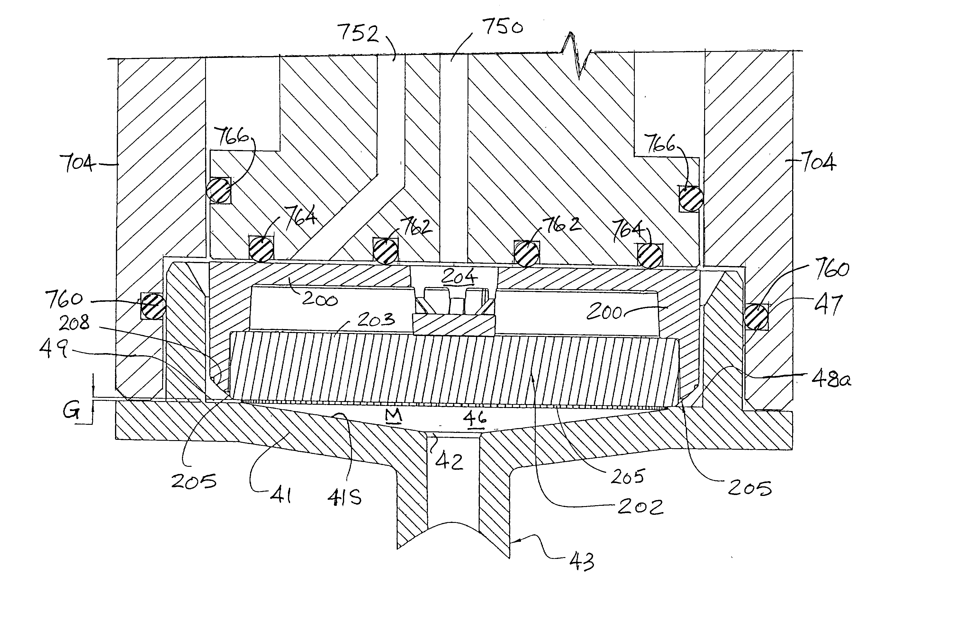
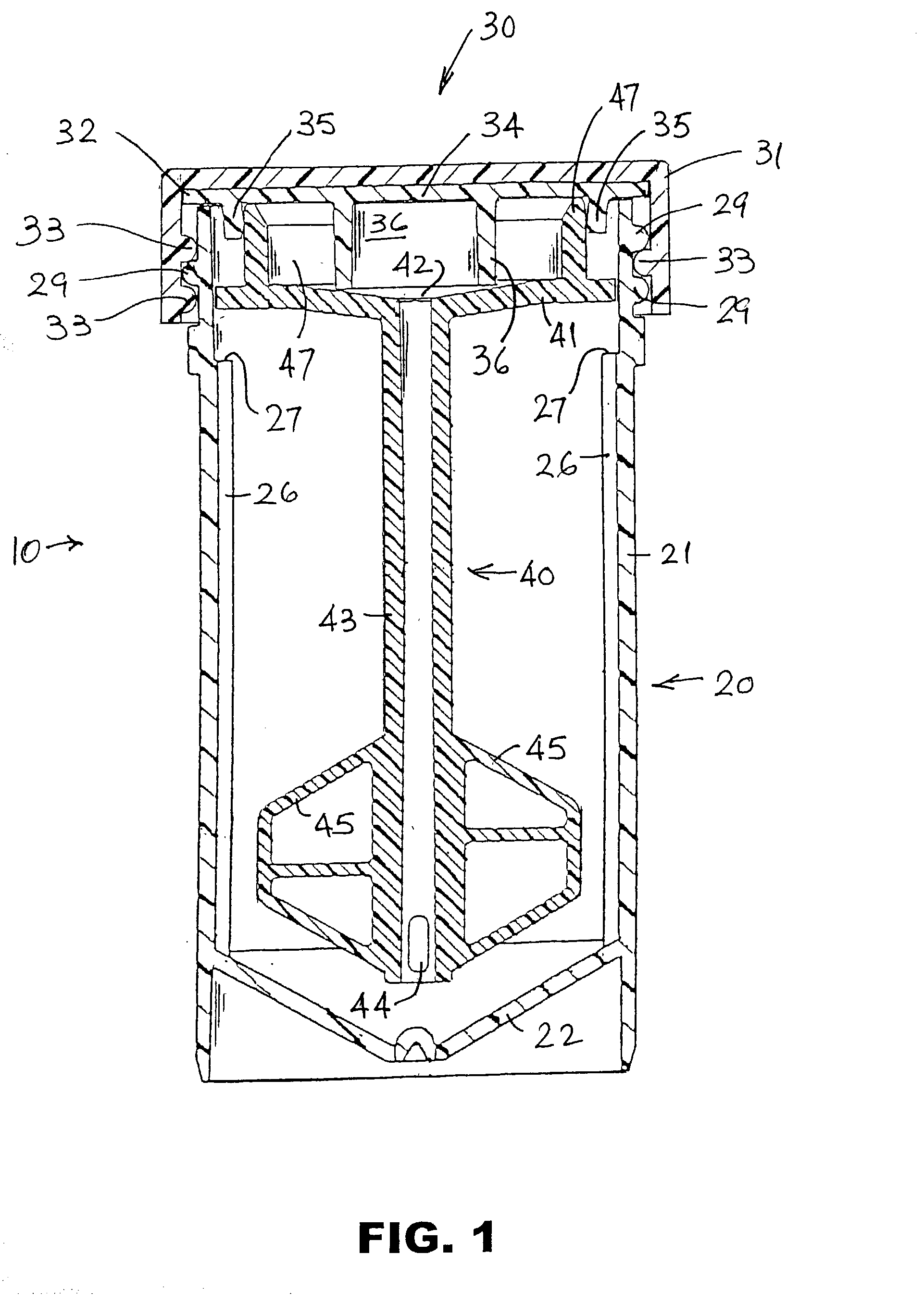
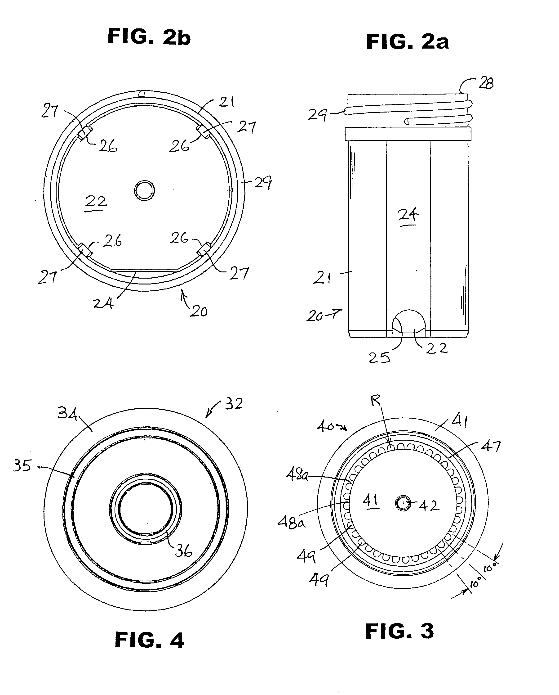
[0092] A full description of this vial-based specimen handling and processing system must begin with the vial itself, which consists of a container, a cover and a processing assembly (stirrer) in the vial.

Specimen Vial

[0093] Referring to FIGS. 1, 2a and 2b, the vial 10 comprises a container 20, a cover 30 and a processing assembly 40. Processing assembly 40 is designed to carry out several functions, among them mixing, and for this preferred rotary embodiment will be referred to as a stirrer for the sake of convenience. Container 20 preferably is molded of a translucent plastic, preferably polypropylene, and has a substantially cylindrical wall 21, surrounding its longitudinal axis, joined to a conical bottom wall 22. Possible alternative plastics include ABS and polycyclohexylenedimethylene terephthalate, glycol (commercially available from Eastman Kodak Co. under the name EASTAR.RTM. DN004). A small portion 24 of wall 21 preferably is flat, the outer surface of the flat portion ad...


PUM

PropertyMeasurementUnit
angleaaaaaaaaaa
heightaaaaaaaaaa
inner diameteraaaaaaaaaa

Abstract

Apparatus and method for handling a container having a removable cover, e.g., a vial for holding a biological fluid specimen. The container may have therein an internal element that is releasably coupled to the cover, such as an assembly for processing the specimen in the container. The method involves applying an external force to the covered container sufficient to release the internal element from the cover, and then dislodging the cover from the container. The external force may be an acceleration force applied to the container, such as an impact force to the bottom of the container, or direct contact with the cover with sufficient force to deflect the cover inwardly. A vertically mounted pressing member and a movably mounted gripper can be used. The pressing member may act to eject the cover from the gripper after the container is clear of the gripper. The apparatus may be part of an automated system for processing multiple liquid-based specimens.

Description

[0001] This application claims the benefit of commonly owned U.S. provisional application No. 60 / 330,092, filed Oct. 19, 2001, No. 60 / 372,080, filed Apr. 15, 2002, and No. 60 / 373,658, filed Apr. 19, 2002, all of which are incorporated herein by reference. This application also is related to commonly owned U.S. non-provisional application Ser. No. 10 / 122,151, filed Apr. 15, 2002, which is also incorporated herein by reference.[0002] The present disclosure is directed to apparatus and methods for collecting and processing specimens of particulate matter-containing liquid, e.g., biological fluid, including collecting and depositing onto a microscope slide or other surface a uniform layer of particulates therefrom (e.g., cells) suitable for examination (e.g., use in cytology protocols).[0003] Diagnostic cytology, particularly in the area of clinical pathology, bases cytological interpretations and diagnoses on examination of cells and other microscopic objects. The accuracy of the scree...

Claims


Application Information

Patent Timeline
Patent Type & Authority Applications(United States)
IPC IPC(8): B01D61/18B01D61/22G01N1/00B01D63/08B01D65/08B01F35/60B01L3/00B01L3/14B01L9/00B01L9/06B65B7/16B65B69/00B67B7/18C12M1/00C12M1/12C12M1/34C12Q1/02G01N1/02G01N1/10G01N1/28G01N1/36G01N27/22G01N33/483G01N35/00G01N35/02G01N35/04G01N35/10
CPCB01D61/18Y10T29/49822B01D63/08B01D65/08B01D2321/02B01D2321/10B01D2321/16B01D2321/2008B01D2321/2016B01D2321/2066B01F7/00583B01F7/1695B01F13/002B01F13/04B01F13/045B01L3/502B01L3/508B01L3/50825B01L9/06B01L9/52B01L2200/025B01L2300/042B01L2300/044B01L2300/046B01L2300/0681B01L2300/0822B01L2300/0851B65B7/161B65B69/00G01N1/2813G01N1/4077G01N27/22G01N35/00G01N35/00029G01N35/0099G01N35/021G01N35/04G01N35/1016G01N2001/1025G01N2001/2826G01N2035/00089G01N2035/00138G01N2035/0405G01N2035/0427G01N2035/0441G01N2035/0443G01N2035/0458G01N2035/0462Y10T436/112499Y10T436/113332Y10T436/11Y10T436/25375Y10T436/255Y10T436/12Y10T29/53039B01D61/22B01F27/13B01F27/88B01F35/605B01F35/60B01F33/5011B67B7/18
Inventor MAYER, WILLIAM J.
Owner HOLOGIC INC
Why Eureka
Try Eureka