Method and apparatus for surgical training

a surgical training and surgical technology, applied in the field of surgical training devices and methods, can solve the problems of limited training to artificial models, inability to reliably mimic the anatomy and the characteristics of the vascular tree and tissue in the human and animal anatomy during live surgery, and hemorrhage, so as to achieve the effect of replicating the experience of skin-to-skin procedures, and training is limited to a few procedures

Inactive Publication Date: 2003-10-02
ABOUD GHAITH +1
View PDF3 Cites 116 Cited by
  • Summary
  • Abstract
  • Description
  • Claims
  • Application Information

AI Technical Summary

Problems solved by technology

At present, training is limited to artificial models that simulate human anatomy, anesthetized live animals and cadavers, but none of them reliably mimic the anatomy and the characteristics of the vascular tree and tissue in the human and animal anatomy during live surgery, in particular, hemorrhage.
Training models using a portion of animal anatomy are known, but even with such a model, training is limited to a few procedures and cannot replicate the experience of "skin-to-skin" procedures, i.e., from opening to closing.
On the other hand, live anesthetized animals do not represent true human anatomy, apart from the ethical considerations involved in the use of live animals in surgical training.
Unfortunately, these are limited to simple techniques and have no relation to the actual anatomy or surgical crises that are encountered by the trainee in live surgery.
The disclosed devices employ simulations of living anatomy and are generally limited to one or a few procedures.
While such models may be valuable in the early stages of training, they are less effective for higher level training in surgical procedures.
Current laboratory training models, while essential for developing surgical skills, are limited in their effectiveness, especially with microvascular procedures.
The alternative is to use live anesthetized animals, which do not represent true human anatomy and have ethical issues with regard to the use of live animals for surgical training.

Method used


Image

  • Method and apparatus for surgical training
  • Method and apparatus for surgical training
  • Method and apparatus for surgical training

Examples


example 2

[0065] Preparation of Colored Fluid and Operating the System

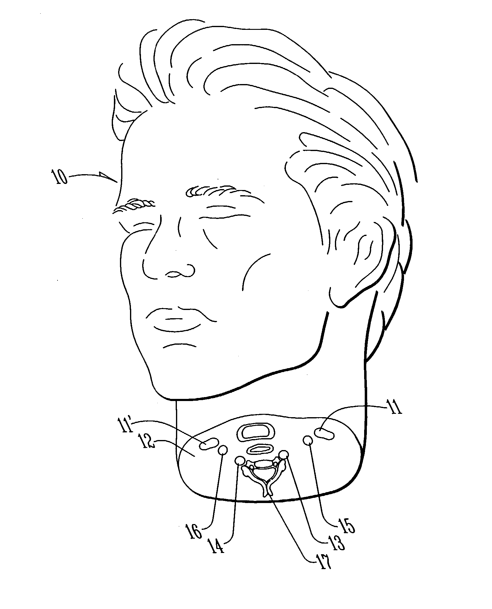
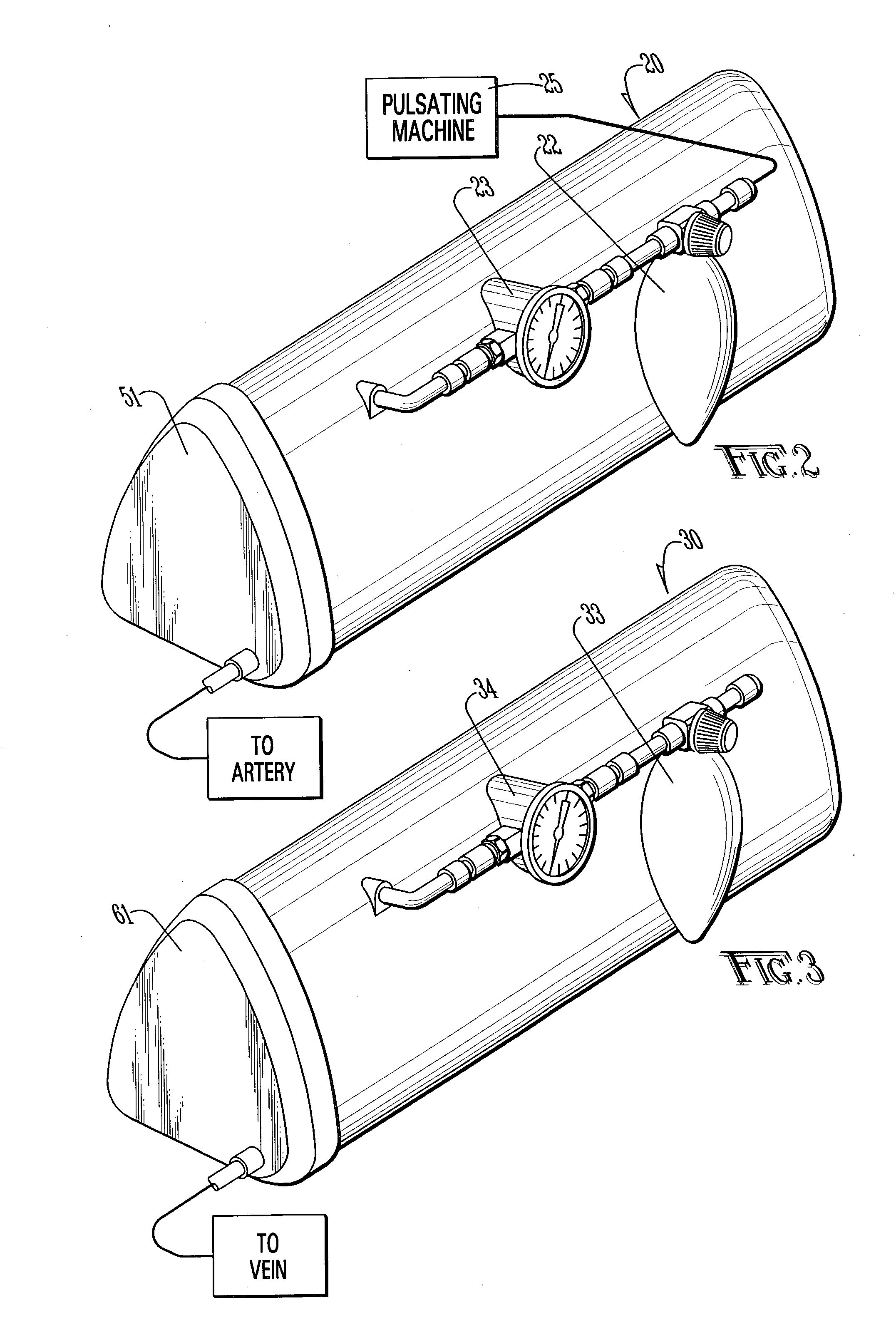
[0066] We used tap water and food coloring to prepare red and dark red fluid. The containers of colored fluid were soft and flexible (serum bags worked well). The arteries were injected with red fluid through the carotid and vertebral arteries on one side until flow appeared on the other side. At this point, we closed the opposite carotid and vertebral arteries and continued injecting both ipsilateral arteries simultaneously, applying moderate pressure to open and fill the terminal branches, then closing the ends. The same was done with the jugular veins.

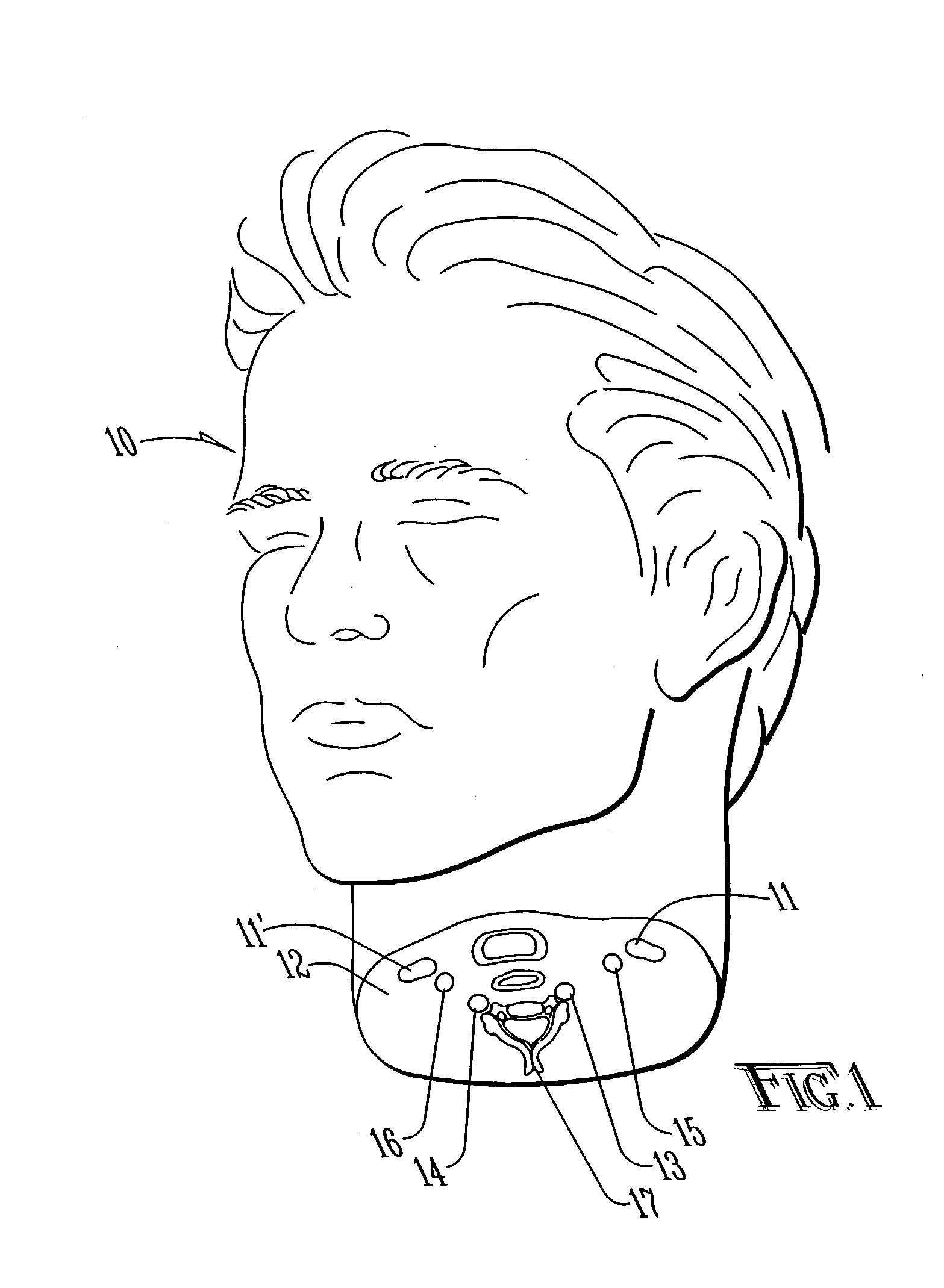
[0067] The pressure bag of the red reservoir was then connected to the pulsating device that provides a pulsating pressure that could be transmitted into the red fluid reservoir through the pressure bag. A rate of 60 pulses per minute was selected. The machine provided a rate of 60 to 120 pulses per minute. Pressure of up to 150 mm Hg could be applied through the pressure b...

example 3

[0070] Craniotomy

[0071] A large scalp flap was made to allow a variety of approaches. The superficial temporal artery (STA) was preserved for practice of the STA-MCA bypass. Any bleeding vessels were ligated, coagulated, or clamped by Raney clips. According to the intended procedure, a variety of craniotomies were performed, with care to preserve the underlying dura. The edges of the bone were waxed to prevent a fluid leak. The dura was opened and leaking vessels were coagulated.

example 4

[0072] Cisternal and Vascular Dissection

[0073] The exposed brain was lifelike, with pulsation and fluid filled cisterns, the arteries were red and pulsating, the veins were darker and filled, and a clear fluid simulated the release of cerebrospinal fluid when the arachnoid was opened. The sylvian fissure was split, the branches of the MCA was followed down to the carotid and basal cisterns, dissecting the branches of the circle of Willis, and exposing all the neurovascular structures in the skull base.


PUM


Abstract

An apparatus and method for microsurgical training using cadaveric anatomy with filling of the vascular system by fluids under pressure to simulate the appearance and function of live surgery. One or more arteries on the specimen of cadaveric anatomy are cannulated and connected to an arterial reservoir having a flexible container holding an arterial fluid simulating the appearance of blood circulating in the arteries of the living organism from which the cadaveric anatomy is derived. Suitable static pressure simulating the arterial pressure appropriate to that of the living organism is applied to the air in an air-tight space surrounding the flexible container in the arterial reservoir. A pulsating machine provides air pulsations to the space surrounding the flexible fluid container to simulate the normal pulsations of the arterial system. One or more veins on the specimen are also cannulated and connected to a venous reservoir having a flexible container holding a venous fluid simulating the appearance of blood circulating in the veins of the living organism. Suitable static pressure simulating the venous pressure appropriate to that of the living organism is applied to the air in an air-tight space surrounding the flexible container in the venous reservoir. Optionally, if the specimen includes at least a portion of spinal canal, a clear fluid reservoir can be connected to the specimen through the spinal canal to simulate cerebrospinal fluid.

Description

[0001] This application claims the benefit of U.S. Provisional Application No. 60 / 368,458 filed Mar. 28, 2002.[0002] Not applicable.[0003] 1. Field of the Invention[0004] The present invention relates to devices and methods for surgical training, and in particular, to such devices and methods which are useful in training on microvascular and microsurgical procedures on both human and animals.[0005] 2. Brief Description of the Related Art[0006] Laboratory training models are essential for developing and refining surgical skills, especially for microsurgery. The closer to live surgery the model is, the greater the benefit. At present, training is limited to artificial models that simulate human anatomy, anesthetized live animals and cadavers, but none of them reliably mimic the anatomy and the characteristics of the vascular tree and tissue in the human and animal anatomy during live surgery, in particular, hemorrhage. Training models using a portion of animal anatomy are known, but e...

Claims


Application Information

Patent Timeline
Patent Type & Authority Applications(United States)
IPC IPC(8): G09B23/28G09B23/30
CPCG09B23/303
Inventor ABOUD, EMAD T.
Owner ABOUD GHAITH
Why Eureka
Try Eureka