
Needle biopsy forceps with integral sample ejector

a biopsy forcep and integral technology, applied in the field of flexible biopsy forceps, can solve the problems of difficult and cumbersome tasks, increased risk for medical personnel from infectious samples, and difficulty in safely removing samples from the forceps needl

Inactive Publication Date: 2005-03-03
GRANIT MEDICAL INNOVATION
View PDF23 Cites 77 Cited by
  • Summary
  • Abstract
  • Description
  • Claims
  • Application Information

AI Technical Summary

Benefits of technology

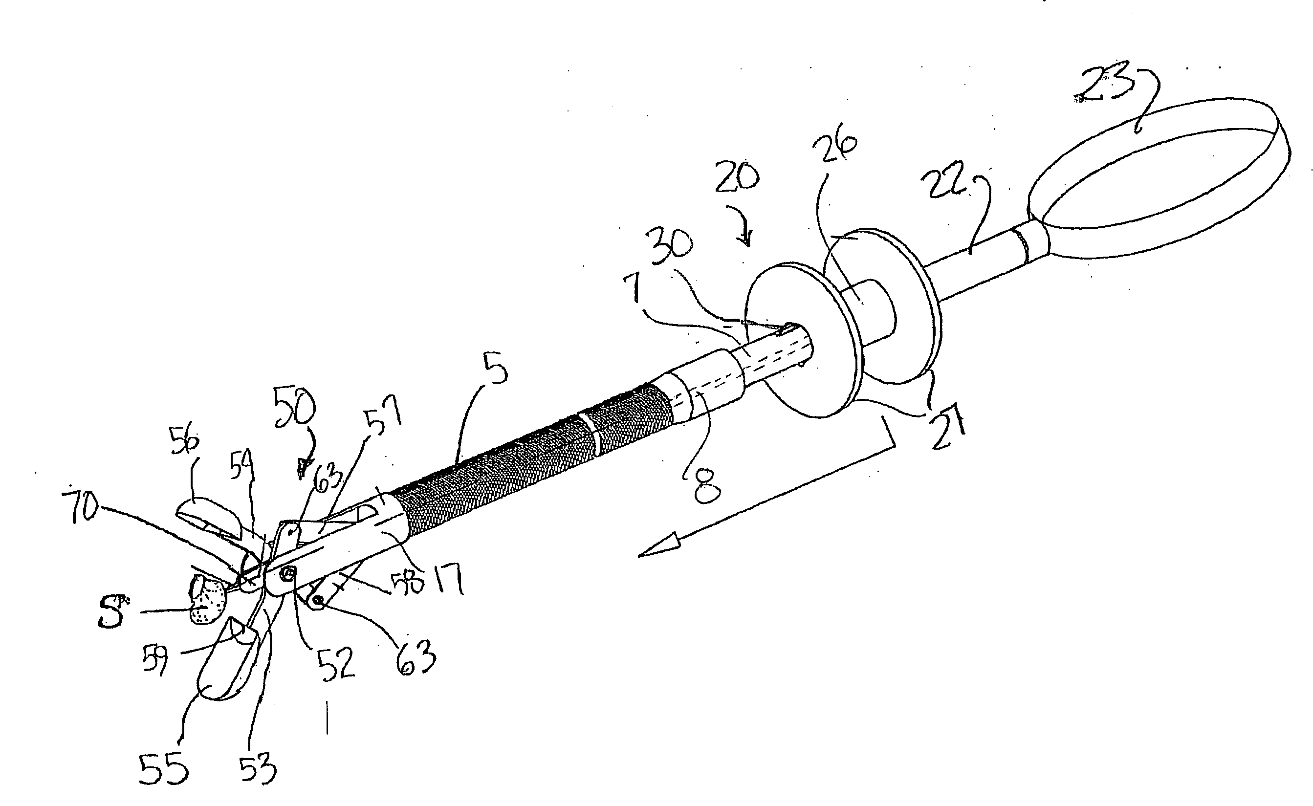
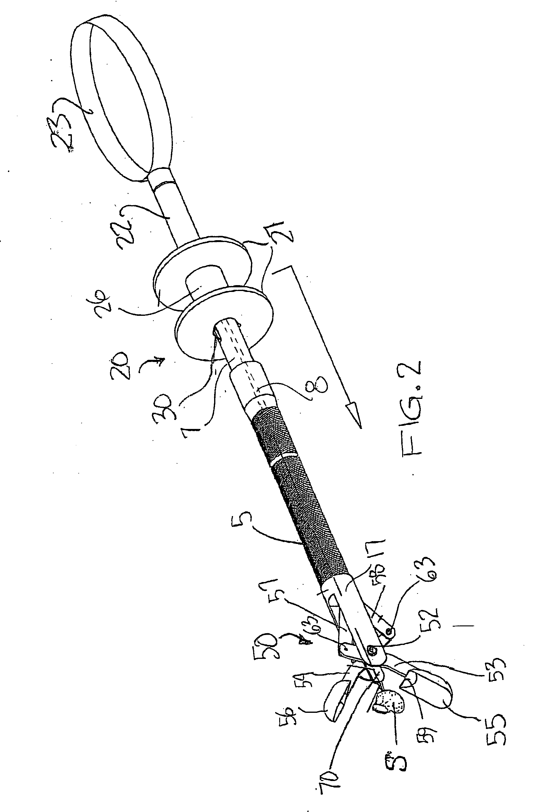
[0011] As broadly contemplated, the sample ejecting safety biopsy needle forceps of the invention comprises a tissue sample contacting surface located at the proximal end of the needle and means for producing relative movement between this surface and the needle to thereby contact the sample and push it along the longitudinal axis of the needle when the cups are in the open position after removal from the hollow cable. This allows the physician or an assistant to safely and precisely deposit the sample in a container for later analysis. The relative movement is accomplished by control means located at the proximal end of the forceps. The control means preferably comprises the handle or, alternatively, is positioned near the handle. At least one linking member is slidably disposed in a hollow cable and extends from the control means in the handle to the proximal end of the instrument.

Problems solved by technology

In many instances, it is difficult to safely remove the sample from the forceps needle.
This task is difficult and cumbersome in itself, and dangerous since there are occasions in which medical personnel have been stuck by the needle.
An additional risk to the medical personnel from an infectious sample is posed by the sharp cutting edges of the cups themselves, which must be maintained in the open position while the tissue sample(s) are removed from the forceps needle.

Method used


Image

  • Needle biopsy forceps with integral sample ejector
  • Needle biopsy forceps with integral sample ejector
  • Needle biopsy forceps with integral sample ejector

Examples


Embodiment Construction

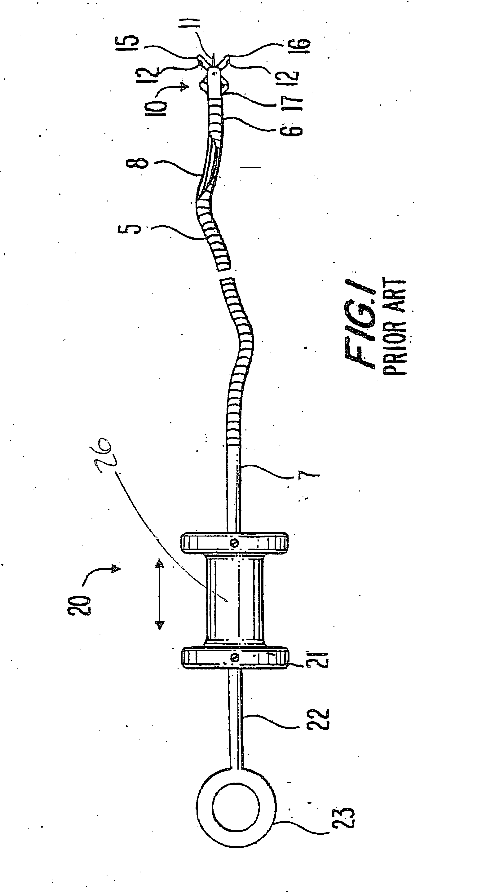
[0053] A conventional needle biopsy forceps 1 of a type known in the art is illustrated in FIG. 1. The forceps comprises a hollow, flexible cable 5 having a distal end 6 and a proximal end 7. An actuating wire 8 is slidably disposed within the cable and extends from the distal to the proximal ends of the cable. At the distal end of the cable is a tissue sample collection means 10, typically comprising two opposing cups 15 and 16 pivotally attached to a clevis 17 depending from the cable 5. A needle or spike 11 extends from the distal end and is enclosed by cups 15 and 16 when they are in the closed position. The cups are operably attached to the wire 8, so that sliding the wire distally opens the cups, and sliding the wire proximally closes the cups.

[0054] With continuing reference to FIG. 1, there is shown at the proximal end 7 of the flexible external cable 5 an actuation handle means 20, typically comprising a spool 26 with flanges 21 slidably mounted on shaft 7. The shaft 22 ha...


PUM


Abstract

A needle biopsy instrument for retrieving a tissue sample from an internal organ such as needle biopsy forceps, includes a pushing surface for safely dislodging a tissue sample from the needle after the instrument containing the sample is retrieved from the patient using mechanical control means operated from the proximal handle to thereby avoid the risk of accidental cuts, punctures and other injuries to medical personnel while trying to manually remove the sample. The instrument has a cam mechanism,or other mechanical links scissor links for operating the cups and producing relative movement between the pushing surface and axis of the needle to cause a tissue sample to be moved toward and eventually off of the tip of the needle into a sample receiving cup. An axial reciprocating control member attached to either the needle or the pushing surface produces the relative movement and is manually controlled by a handle or other grip at the proximal end of the forceps. The needle is either retracted axially from the distal toward the proximal end of the device, or a pushing member attached to the distal end of an actuating wire or cable is moved distally along the axis of a stationary needle. A latch mechanism can be provided on the proximal end to release a biasing force to provide the relative movement between the needle and pushing member to slide a tissue sample off the needle.

Description

FIELD OF THE INVENTION [0001] The invention relates to flexible biopsy forceps used in conjunction with an endoscope and rigid forceps used with a laparoscope for retrieval of a tissue sample from the interior of a patient's body, where the forceps include a needle that passes through the tissue to be sampled in order to retain one or more severed samples for retrieval. BACKGROUND OF THE INVENTION [0002] Flexible needle biopsy forceps are used in conjunction with a fiber optic endoscope as follows: the endoscope is inserted into the stomach, colon or other hollow organ of the patient's body, an abnormality is visualized, and the flexible biopsy forceps is introduced through the biopsy channel of the endoscope. The distal end of the biopsy forceps is comprised of two opposed sharp-edged cups that are operably attached by means of pivot arms to a wire passing on the interior of a flexible cable. A fixed spike or needle is positioned within the closed cups. As used hereinafter, the ter...

Claims


Application Information

Patent Timeline
Patent Type & Authority Applications(United States)
IPC IPC(8): A61B10/00A61B10/06A61B17/28
CPCA61B10/06A61B17/29A61B2017/2939A61B2017/2926A61B2017/2911
Inventor NAKAO, NAOMI L.
Owner GRANIT MEDICAL INNOVATION
Learn More