Radiation tomograph apparatus and radiation tomography method thereof

a tomograph and radiation tomography technology, applied in the field of tomograph apparatus and radiation tomography method thereof, can solve the problems of reducing the contrast of tomographic images and artifacts, deteriorating image quality, and difficult to obtain generated amounts of crosstalk in the body axial direction, so as to prevent the reduction of tomographic image contrast and improve the quality of tomographic images

Inactive Publication Date: 2005-03-10
GE MEDICAL SYST GLOBAL TECH CO LLC
View PDF11 Cites 10 Cited by
  • Summary
  • Abstract
  • Description
  • Claims
  • Application Information

AI Technical Summary

Benefits of technology

[0015] According to the present invention, a radiation tomography apparatus capable of preventing a reduction in the contrast of a tomographic image due

Problems solved by technology

Therefore, when a tomographic image is reconstructed and generated based on detection data containing the crosstalk, there might be cases where in the X-ray CT apparatus, a reduction in the contrast of a tomographic image and artifacts occurred and the quality of an image was deteriorated.
However, it was difficult to obtain the generated amounts of crosstalk in the body axial direction.
Therefore, the prior art encoun

Method used


Image

  • Radiation tomograph apparatus and radiation tomography method thereof
  • Radiation tomograph apparatus and radiation tomography method thereof
  • Radiation tomograph apparatus and radiation tomography method thereof

Examples


embodiment 1

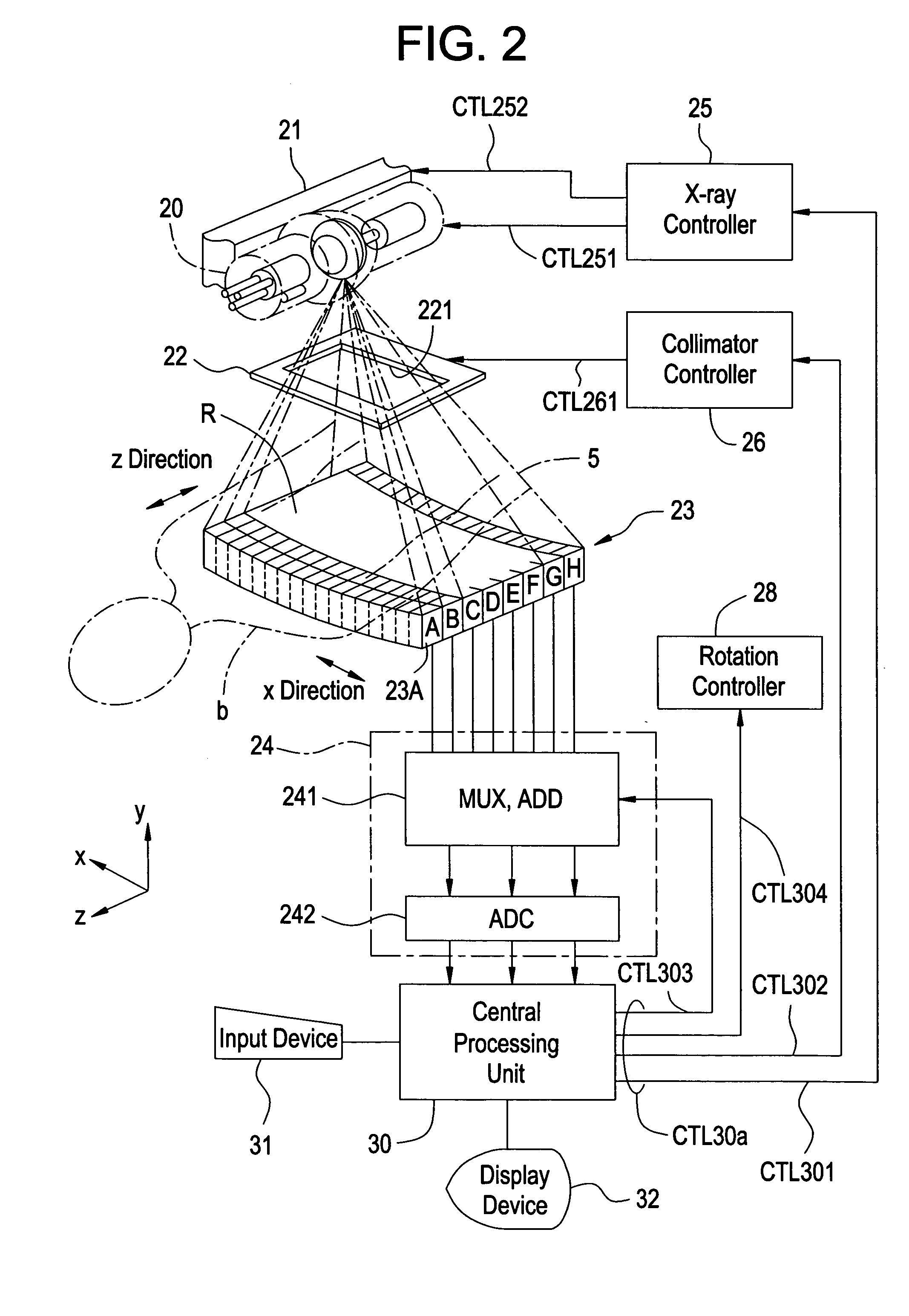
[0027]FIG. 1 is a block diagram showing an overall configuration of an X-ray CT apparatus 1 used as a radiation tomography apparatus according to an embodiment 1 of the present invention. FIG. 2 is a configurational diagram illustrating an essential part of the X-ray CT apparatus 1 used as the radiation tomography apparatus according to the embodiment 1 of the present invention.

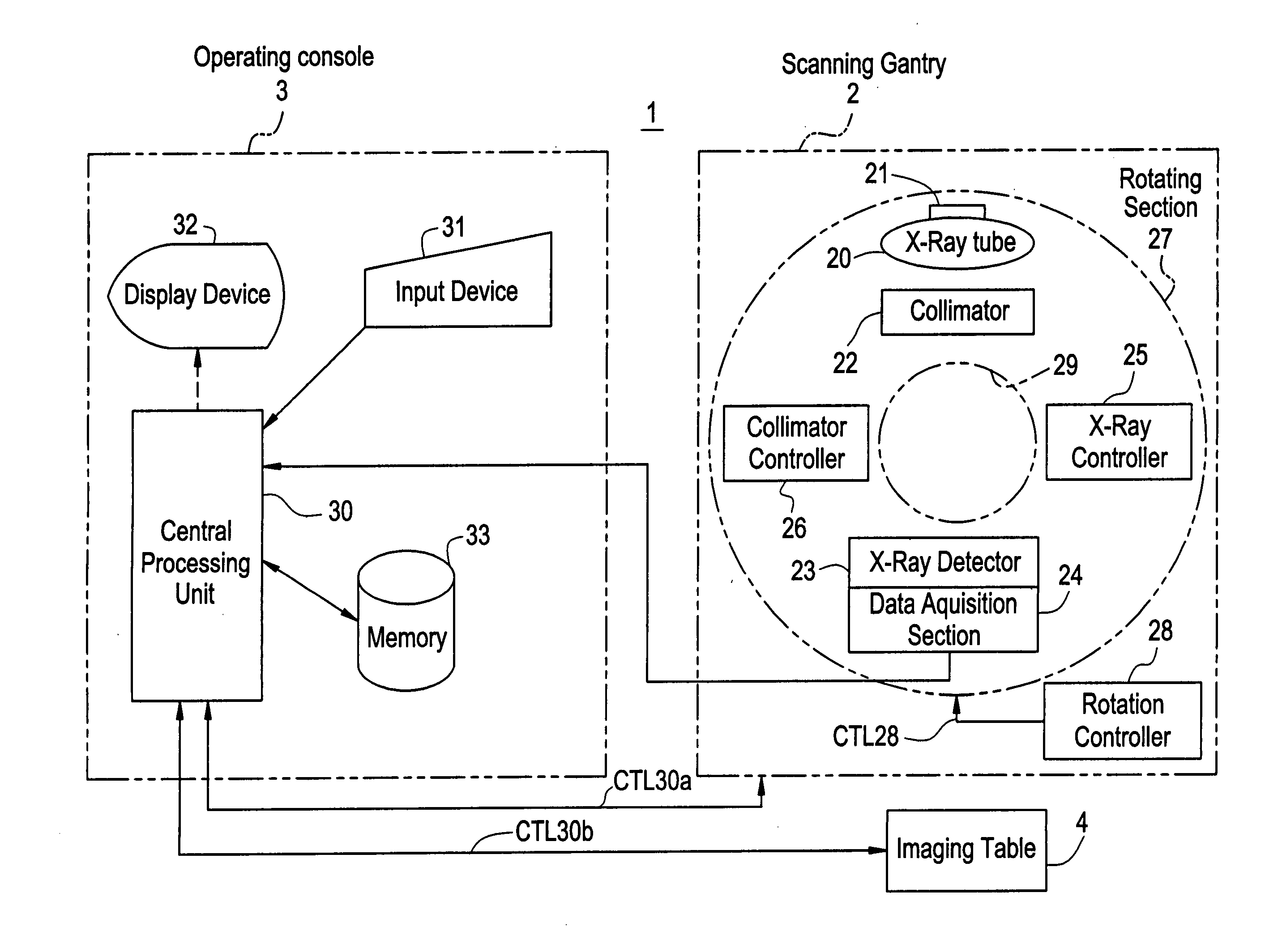
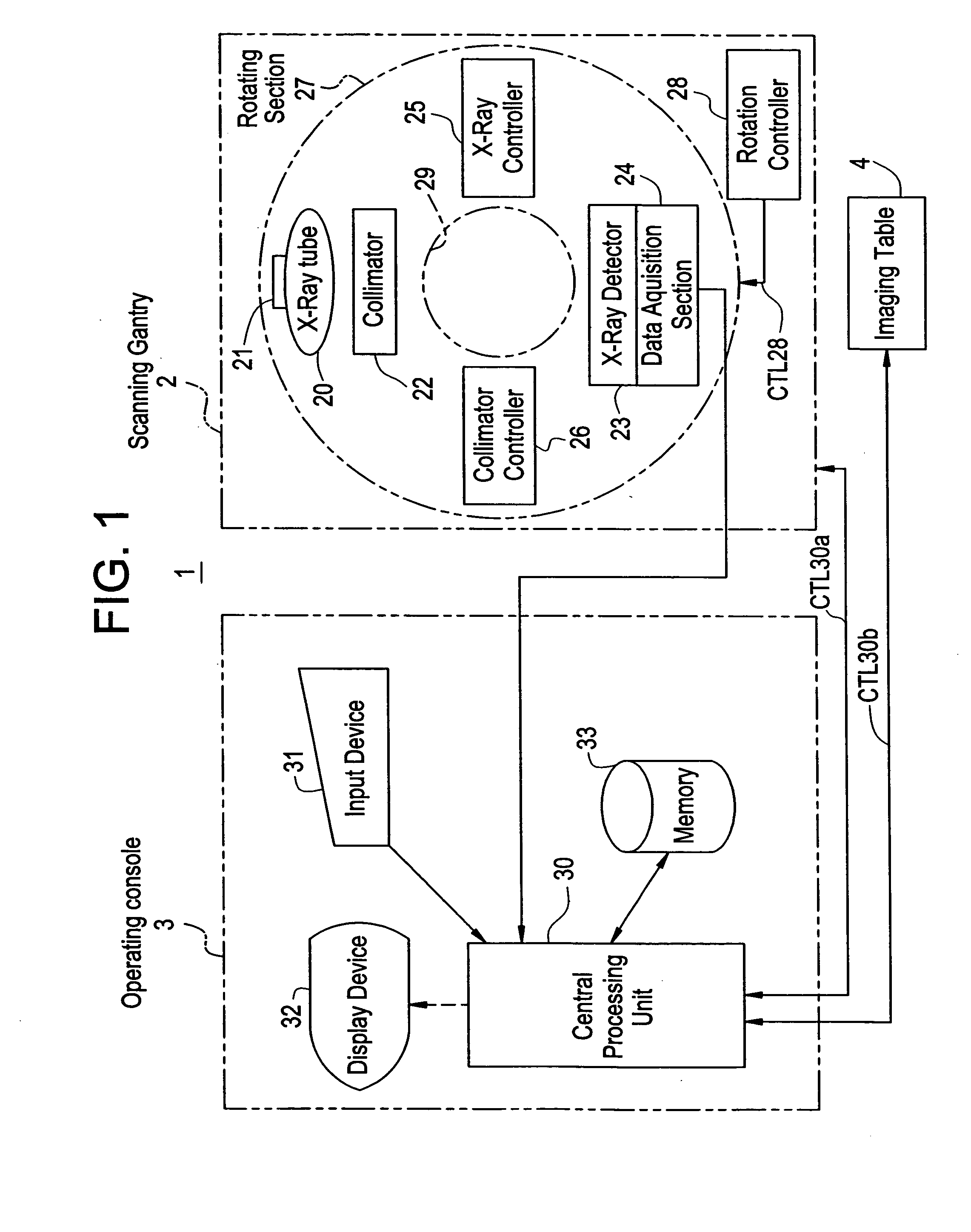
[0028] As shown in FIG. 1, the X-ray CT apparatus 1 according to the present embodiment includes a scanning gantry 2, an operating console 3 and an imaging table 4.

[0029] The scanning gantry 2 includes an X-ray tube 20, an X-ray tube moving section 21, a collimator 22, an X-ray detector array 23, a data acquisition section 24, an X-ray controller 25, a collimator controller 26, a rotating section 27 and a rotation controller 28 as principal components. Here, the X-ray tube 20 and the X-ray detector array 23 are opposed to each other with an X-ray irradiation space 29 interposed therebetween.

[0030] Incident...

embodiment 2

[0072] An X-ray CT apparatus used as a radiation tomography apparatus according to an embodiment 2 of the present invention has such configurations as shown in FIGS. 1 and 2 in a manner similar to the embodiment 1. The X-ray CT apparatus according to the present embodiment is similar to the embodiment 1 except that a fitting processing means is different from that employed in the embodiment 1. Therefore, the description of parts common to the X-ray CT apparatus according to the embodiment 1 will be omitted.

[0073] The fitting processing means of the X-ray CT apparatus according to the present embodiment is provided in a central processing unit 30 in a manner similar to the embodiment 1. In the present embodiment, the fitting processing means calculates dummy data corresponding to first detection data according to fitting processing using only second detection data and substitutes the dummy data with the first detection data.

[0074] An X-ray tomography method using the X-ray CT appar...

embodiment 3

[0078] An X-ray CT apparatus used as a radiation tomography apparatus according to an embodiment 3 of the present invention has such configurations as shown in FIGS. 1 and 2 in a manner similar to the embodiments 1 and 2. The X-ray CT apparatus according to the present embodiment is similar to the embodiments 1 and 2 except that a fitting processing means is different from ones employed the embodiments 1 and 2. Therefore, the description of parts common to the X-ray CT apparatuses according to the embodiments 1 and 2 will be omitted.

[0079] The fitting processing means of the X-ray CT apparatus according to the present embodiment is provided in a central processing unit 30 in a manner similar to the embodiments 1 and 2. In the present embodiment, the fitting processing means calculates first dummy data corresponding to first detection data according to fitting processing using second detection data by means of a plurality of X-ray detection modules respectively and calculates differ...


PUM


Abstract

A method and apparatus for reducing the contrast of a tomographic image due to crosstalk and the occurrence of artifacts and improve the quality of the tomographic image. Detection data are subjected to fitting processing in such a manner that, of the data generated by an X-ray detected by X-ray detection elements disposed in array form in a plurality of X-ray detection modules disposed adjacent to one another, first detection data generated by the X-ray detected by X-ray detection elements adjacent to a boundary between the plurality of X-ray detection modules are adapted to waveform data based on second detection data generated by the X-ray detected by X-ray detection elements lying around the adjacent X-ray detection elements.

Description

BACKGROUND OF THE INVENTION [0001] The present invention relates to a radiation tomography apparatus and a radiation tomography method thereof. [0002] An X-ray CT (Computed Tomography) apparatus that generates an image of a tomographic plane of a subject to be imaged using an X-ray corresponding to radiation has been known as a radiation tomography apparatus. The X-ray CT apparatus has been used in widespread applications such as medical and industrial applications with a human body and an object as subjects to be imaged. [0003] The X-ray CT apparatus scans the periphery of a subject to be imaged with a subject's body axis direction as the axis and applies X-rays to the subject from a plurality of view directions through an X-ray tube. The X-rays radiated from the X-ray tube are blocked or cut off thereby forming an X-ray so as to be applied to an imaging area of the subject. The X-rays transmitted through the subject from the plural view directions via the collimator are detected b...

Claims


Application Information

Patent Timeline
IPC IPC(8): G01T1/00A61B5/03A61B6/03G01N23/04G01T1/161H04N5/32
CPCA61B6/032G01N2223/612G01N2223/419G01N23/046A61B6/03
Inventor GOHNO, MAKOTONUKUI, MASATAKE
Owner GE MEDICAL SYST GLOBAL TECH CO LLC
Why Eureka
Try Eureka