
Apparatus for assembling anterior cruciate ligament reconstruction system

a technology for assembling and repairing anterior cruciate ligaments, applied in the field of orthopaedic surgery, can solve the problems of poor healing effect, extensive morbidity, and further damage to menisci and articular cartilag

Inactive Publication Date: 2005-03-24
COAPT SYST INC
View PDF99 Cites 203 Cited by
  • Summary
  • Abstract
  • Description
  • Claims
  • Application Information

AI Technical Summary

Benefits of technology

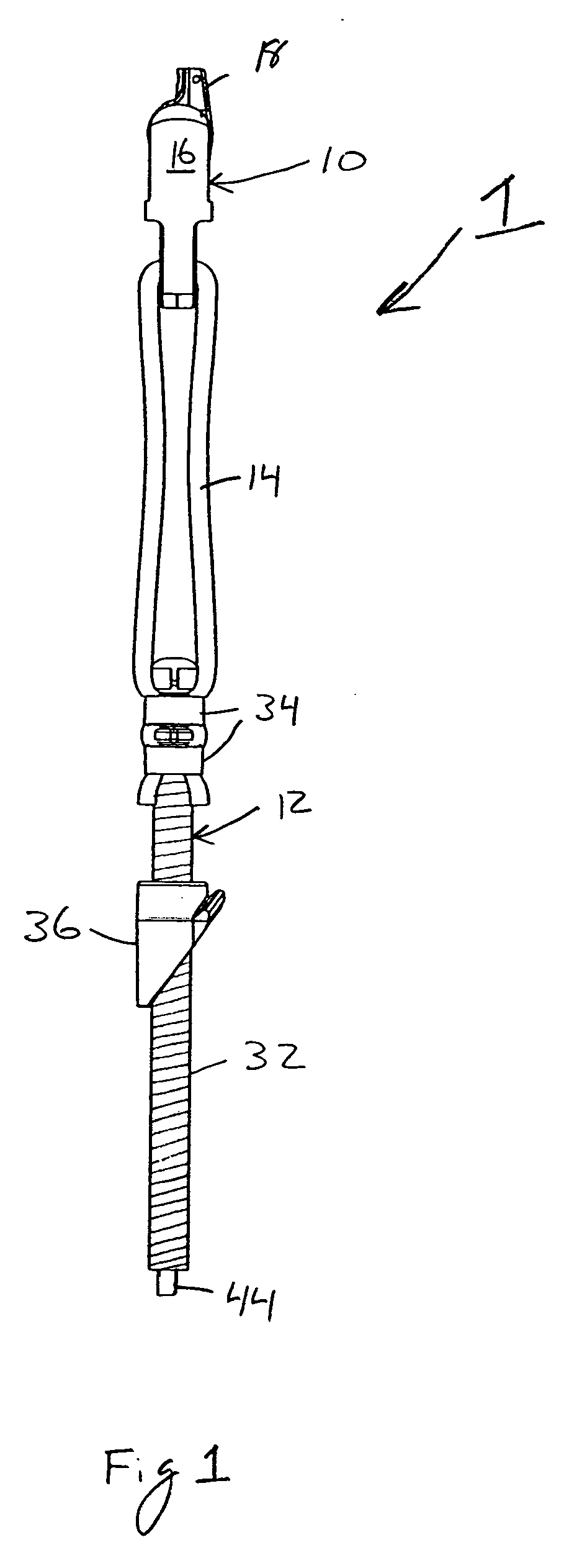
[0020] In still one more aspect of the present invention, an apparatus for mounting a graft to an anchor assembly of a reconstruction system includes a base plate, a first mounting block mounted to the base plate and having a first reference surface, a second mounting block mounted to the base plate and having a second reference surface against which the anchor assembly is mountable that is adjustably moveable relative to the first reference surface, and a tension apparatus mounted to the base plate for applying a tension to a graft connected to the first mounting block, and for positioning the graft to extend adjacent to and past the second mounting block under the tension.
[0021] One more aspect of the present invention is a method for assembling a reconstruction system for implementation into a bone tunnel, wherein the reconstruction system includes a first anchor assembly having a first tissue presentation surface and a second anchor assembly having a tissue fixation surface and a second tissue presentation surface. The method includes mounting the first anchor assembly against a first reference surface of a first mounting block, mounting the second anchor assembly against a second reference surface of a second mounting block, connecting a graft to the ...

Problems solved by technology

The ACL has poor healing properties, and thus, an untreated injury potentially leads to recurrent “giving-way” episodes, further damage to the menisci and articular cartilage, and possible progression to osteoarthritis (Brown et al., Clinics in Sports Medicine 18(1): 109-170 (1999)).
Although the use of a bone-tendon-bone graft may provide the advantage of effective healing due to the efficient biointegration of the bone graft to the bone host, the harvesting of a bone-tendon-bone graft typically results in extensive morbidity to the donor knee joint, thus lengthening the patient's resumption of normal physical activity.
On the other hand, it has historically been more difficult to effectuate and maintain accurate fixation of such grafts throughout the healing period where high-tension forces of the knee may act to disrupt the graft construct (e.g. via fixation device slippage or graft failure).
Unfortunately, many existing procedures have proven inadequate for immediately restoring adequate strength and stability to the involved joint.
Furthermore, even if immediate achievement of knee stability is achieved, many current methods are ineffective at maintaining such stability throughout the period when the mechanical phase of graft fixation is ultimately superceded by a permanent biological phase of graft integration.
One difficulty in effectively implanting a fully effective ligament reconstruction is the surgeon's need to balance a number of variables leading to “trade-offs”.
Many ACL reconstruction systems and techniques allow the tension to be set during insertion of the graft, but not subsequent to tissue fixation and bone anchoring, and especially not subsequent to the knee being subjected to its range of motion.
Thus, the final intra-operative resting tension on the graft ligament is either unknown or unadjustable.
If it is determined after tissue fixation and bone anchoring (and possibly after the knee is moved through its range of motion) that the desired ligament tension was not achieved, most ACL reconstruction systems and techniques offer little or no corrective options.
Moreover, anchor structures, such as those in Johnson (U.S. Pat. No. 5,562,668), are complex, bulky, and difficult to use properly.
However, such external anchoring presents several problems.
The presence of a longer segment of stretchable graft within the bone tunnel can have the “bungee cord effect” that can widen the tunnel, impede healing, and damage the graft.
Also, the lack of immobilization of the graft at the articular orifice can lead to lateral motion (windshield or sundial effect), widening of the orifice, impeded healing, and damage to the graft.
Anchoring the graft within the bone tunnel can overcome the problems of external anchoring, but can diminish the strength of the graft anchor since the bone interior is softer and provides an inferior anchoring point.
Internal anchoring typically requires the use of devices that are destructive of the soft graft tissue (as described below).
Finally, anchoring the ligaments entirely within the bone tunnel precludes the surgeon from properly adjusting the tension on the graft after it has been placed within the tunnel.
However, such devices may create a gap between the bone and the ligament graft, thereby precluding maximal graft-tunnel contact at the point of immobilization, thus possibly impeding healing.
Such constructs require a continuous high-pressure load against both the graft and the surrounding bone, thus possibly leading to damage to the graft and erosion of the bone.
Impeded healing or loosening of the interference fixation, and thus loss of fixation and graft slippage, can often result.
Such an outcome could represent a failure of the operative procedure.
Lastly, healing can be impeded because there is no separation between the fixation and healing portions of the graft.
Tissue necrosis at the tissue fixation portion of the graft can impede healing to the adjacent bone.
These procedures, however, typically require the surgeon to use a graft having a length such that it extends beyond the cortex of the bone tunnel, and bends at approximately a 90 degree angle so that the graft end is flush against the external bone surface for securing to the external bone, which is not ideal.
Stainless steel staples, buttons with sutures, and other related fixation devices have each been used for external anchoring, with limited success, because external fixation devices can have a high profile, are uncomfortable for the patient during healing, and can require a second surgery to remove them.

Method used


Image

  • Apparatus for assembling anterior cruciate ligament reconstruction system
  • Apparatus for assembling anterior cruciate ligament reconstruction system
  • Apparatus for assembling anterior cruciate ligament reconstruction system

Examples


first alternate embodiment

[0090] First Alternate Embodiment of Reconstruction System

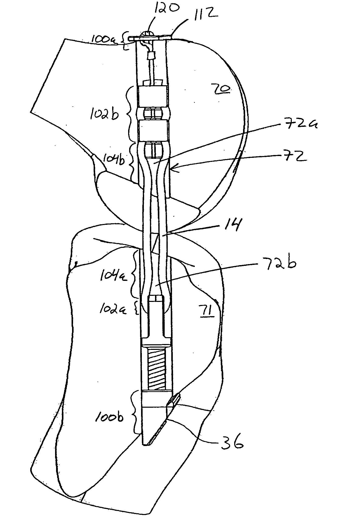
[0091] FIGS. 14, 15A-15D, 16, 17A-17B and 18 illustrate a first alternate embodiment of the reconstruction system of the present invention, where the fixation of the graft using an opening for graft looping is on the tibial assembly, and the fixation of the graft using the compression ring and graft fixation surface is on the femoral assembly. This embodiment includes a femoral assembly 106 and a tibial assembly 108, with a graft 14 spanning therebetween, as best shown in FIG. 14.

[0092] Femur assembly 106 is best shown in FIGS. 15A-15D, and includes a bolt 110 and an anchor plate 112 (bone anchor) rotatably (pivotally) connected thereto. Bolt 110 includes a shaft 114, with a bolt head 116 and a flange 118 at one end thereof (which correspond to the bolt head 42, the flange 46 and the tissue / graft fixation surface described above). The other end of the shaft 114 terminates in an open (e.g. hook shaped) or a closed (e.g. a rin...


PUM

PropertyMeasurementUnit
Lengthaaaaaaaaaa
Forceaaaaaaaaaa
Distanceaaaaaaaaaa

Abstract

An apparatus and method for assembling a soft tissue reconstruction system having first and second anchor assemblies. The apparatus includes a base plate, a first mounting block mounted to the base plate and having a first reference surface against which the first anchor assembly is mountable, a second mounting block slidably mounted to the base plate and having a second reference surface against which a bone anchor of the second anchor assembly is mountable, a measurement bar extending from the second mounting block and slidable relative to the first mounting block, a support block slidably mounted to the measurement bar having a third reference surface for abutting against a tissue presentation surface of the second anchor assembly, and a tensioning device for tensioning a graft attached to the first anchor assembly. Indicia on the measurement bar indicate the proper locations of the reference surfaces and components mounted / abutted thereto.

Description

[0001] This application is a continuation-in-part of U.S. application Ser. No. 10 / 701,917, filed Nov. 4, 2003, which is a continuation-in-part of U.S. application Ser. No. 10 / 159,513, filed May 31, 2002, which claims the benefit of U.S. Provisional Application No. 60 / 295,389, filed May 31, 2001; and also claims the benefit of U.S. Provisional Application No. 60 / 445,259, filed Feb. 4, 2003.FIELD OF THE INVENTION [0002] The present invention relates to the field of orthopedic surgery, and more particularly to an apparatus for assembling ex-vivo an orthopedic surgical device or system that is used to reconstruct soft tissue, such as tendons and ligaments, within the knee or other parts of the body. BACKGROUND OF THE INVENTION [0003] The present invention is primarily directed to the reconstruction of the anterior cruciate ligament (ACL) of the knee. The ACL is a two-bundle ligament that helps to stabilize the knee joint, and prevents posterior displacement of the femur on the tibia and...

Claims


Application Information

Patent Timeline
IPC IPC(8): A61F2/00A61F2/02A61F2/08
CPCA61F2/08A61F2310/00179A61F2/0811A61F2002/0829A61F2002/0852A61F2002/0882A61F2210/0004A61F2240/001A61F2240/005A61F2250/0007A61F2250/0012A61F2310/00017A61F2310/00023A61F2310/00071A61F2/0805
Inventor MAGEN, HUGH E.ELSON, ROBERTSENATORI, MARK
Owner COAPT SYST INC
Learn More