
Medical image processing apparatus

a technology of image processing and medical imaging, applied in image enhancement, measurement using nmr, instruments, etc., can solve the problems of low detection accuracy of abnormal shadow candidates, quantum noise increases, and the contrast becomes extremely low

Inactive Publication Date: 2005-03-31
KONICA MINOLTA MEDICAL & GRAPHICS INC
View PDF6 Cites 17 Cited by
  • Summary
  • Abstract
  • Description
  • Claims
  • Application Information

AI Technical Summary

Benefits of technology

[0021] According to the above-mentioned apparatus, a detection result is displayed so that the display of a detection result is switchable between before

Problems solved by technology

However, if different types of shadow are overlapped in an image, for example, clustered-microcalcification shadow exists within mass shadow, since clustered-microcalcification shadow within mass shadow has little absorption difference of X rays compared to the case that the shadow exists in shadow of normal tissue such as a fat region or a mammary gland region, its contrast becomes extremely low.
Further, in a region where diffe

Method used


Image

  • Medical image processing apparatus
  • Medical image processing apparatus
  • Medical image processing apparatus

Examples


Embodiment Construction

[0033] Hereinafter, an embodiment of the present invention will be described with reference to figures.

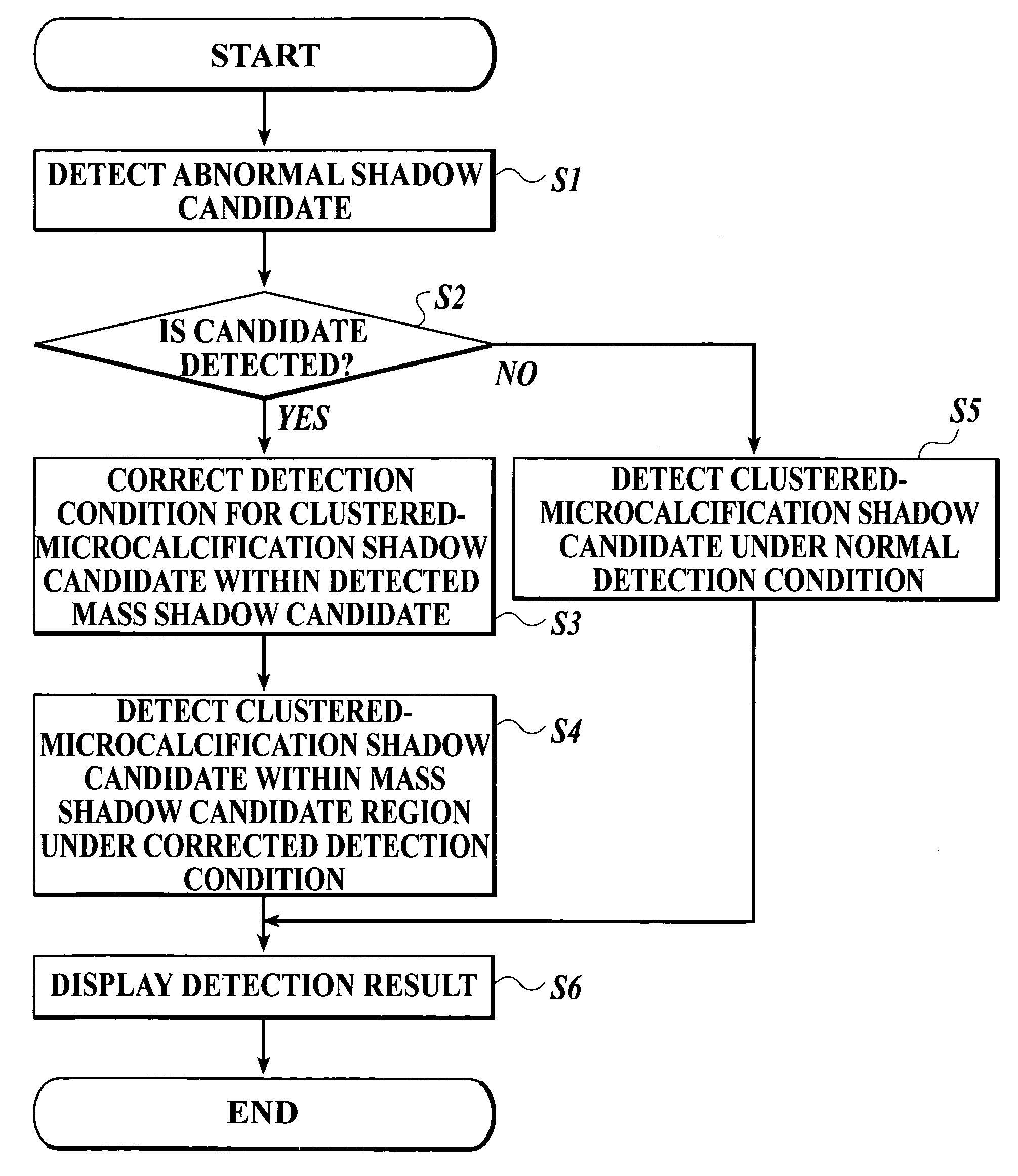
[0034] In the present embodiment, the following example will be described: a detection of a mass shadow candidate or a clustered-microcalcification shadow is performed in an X-ray image in which a mamma of a patient is generated or radiographed (such image is called mammography) according to the finding of breast cancer. Then, in a region where a mass shadow candidate is detected, the detection of clustered-microcalcification shadow is performed under a corrected detection condition for a clustered-microcalcification shadow candidate, for correcting a detection result of a clustered-microcalcification shadow candidate. Here, in the present embodiment, the description will be made with a mammography used as an object. However, the present invention can be applied to a case of detecting abnormal shadow candidates of various types within a medical image of another type, such as an ul...


PUM


Abstract

A medical image processing apparatus includes: an abnormal shadow candidate detecting section for detecting a plurality of types of abnormal shadow candidates in a medical image; and a correcting section for correcting a detection result of one type of abnormal shadow candidate by the abnormal shadow candidate detecting section based on a detection result of another type of abnormal shadow candidate.

Description

BACKGROUND OF THE INVENTION [0001] 1. Field of the Invention [0002] The invention relates to a medical image processing apparatus for detecting a candidate region of abnormal shadow by doing the image analysis on a medical image. [0003] 2. Description of Related Art [0004] In a medical field, there is an occasion where a doctor interprets a medical image such as an X-ray image, an ultrasound image or the like, to find a lesion or to observe a course of a medical condition. So far, for the purpose of reducing burdens on doctor's interpretation of a medical image, the medical image processing apparatus called Computed-Aided Diagnosis (hereinafter, referred to as a CAD) which automatically detects shadow of a lesion as an abnormal shadow candidate by doing the image analysis on image data of the medical image is developed. [0005] For the above-described CAD, a variety of algorithms are developed according to a type of abnormal shadow. For example, as a cancerous part of breast cancer, ...

Claims


Application Information

Patent Timeline
IPC IPC(8): A61B5/00G01R33/54A61B5/055A61B6/00A61B8/08G06K9/00G06T1/00G06T7/00
CPCG06T2207/30068G06T7/0012
Inventor KASAI, SATOSHI
Owner KONICA MINOLTA MEDICAL & GRAPHICS INC
Features
  • Generate Ideas
  • Intellectual Property
  • Life Sciences
  • Materials
  • Tech Scout
  • Unparalleled Data Quality
  • Higher Quality Content
  • 60% Fewer Hallucinations
Learn More