
Biopsy cavity marking device and method

a marking device and cavity technology, applied in the field of subcutaneous cavity marking devices and methods, can solve the problems of clip detachment from the cavity wall, device has significant drawbacks, initial biopsy failed to remove a sufficient amount of lesion, etc., and achieve the effect of minimizing any “dimpling effect”

Inactive Publication Date: 2005-04-14
DEVICOR MEDICAL PROD
View PDF99 Cites 63 Cited by
  • Summary
  • Abstract
  • Description
  • Claims
  • Application Information

AI Technical Summary

Benefits of technology

The device effectively marks the biopsy cavity center and periphery, ensuring accurate follow-up examinations and minimizing the risk of cancer cell spread, with enhanced visibility through non-invasive imaging and reduced interference with healing or re-excision procedures.

Problems solved by technology

In some cases, however, the physician may be concerned that the initial biopsy failed to remove a sufficient amount of the lesion.
This device has significant drawbacks.
If the limbs have been prematurely bent together, the clip will be discarded since it will most likely not attach properly to the cavity wall.
Additionally, there is always the possibility that the clip may detach from the cavity wall during or after withdrawal of the tools used to place the clip into the cavity.
Aside from the problems inherent in the placement of the marking clip, there are also limitations associated with how well the marking clip can identify a biopsy cavity.
Moreover, patient concern limits the number of clips that may be placed in a cavity.
Obviously, determination of the periphery of a biopsy cavity from one point of the periphery is not possible.
These limitations are compounded as the biopsy cavity fills within a few hours with bodily fluids, which eventually renders the cavity invisible to non-invasive techniques.
Another difficulty in viewing the clip stems from the fact that the clip is attached to the side, not the center, of the cavity.
This makes determining the spatial orientation and position of the cavity difficult if not impossible during follow-up examination.
Additionally, during a stereotactic breast biopsy procedure, the breast is under compression when the marking clip is placed.
Upon release of the compressive force, determining the location of the clip can be unpredictable, and the orientation as well as the location of the periphery of the cavity are lost.

Method used


Image

  • Biopsy cavity marking device and method
  • Biopsy cavity marking device and method
  • Biopsy cavity marking device and method

Examples


Embodiment Construction

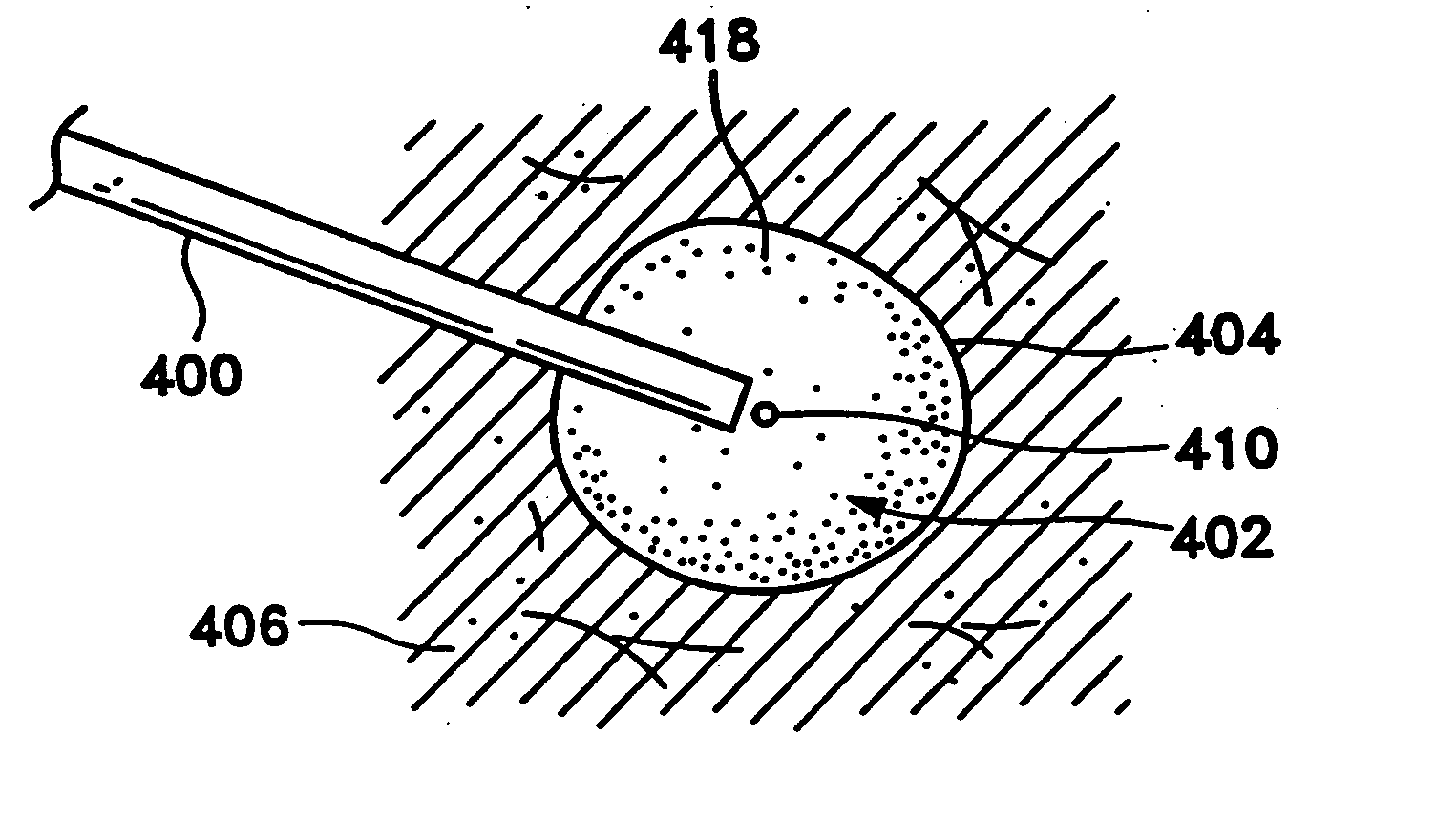
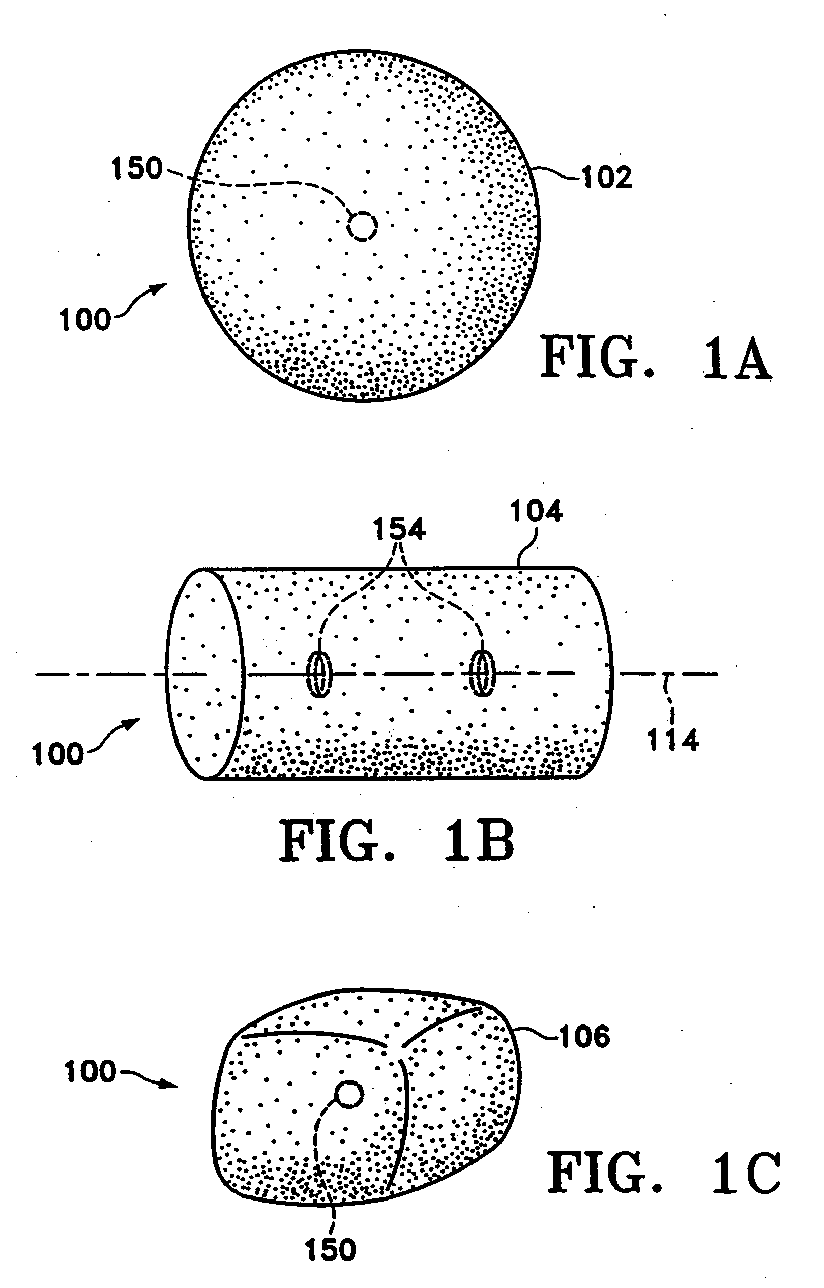
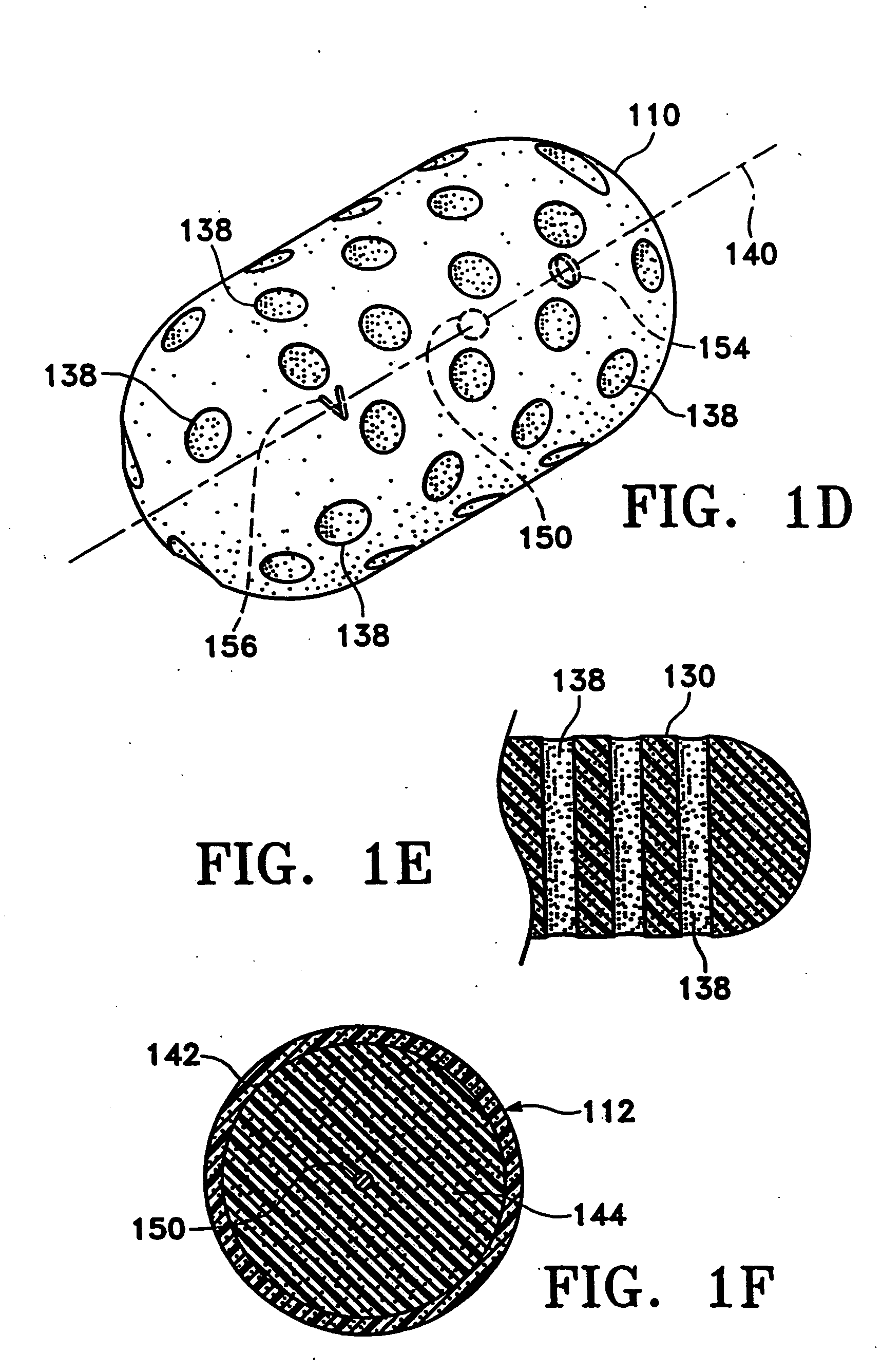
[0048]FIGS. 1A-1C show various configurations of a preferred subcutaneous cavity marking device of the present invention. Here the marking device (100) is displayed as having either a generally spherical body (102) (FIG. 1A), a generally cylindrical body (104) (FIG. 1B), or a multi-faced or irregular body (106) (FIG. 1C). In general, it is within the scope of this invention for the body to assume a variety of shapes. For example, the body may be constructed to have substantially curved surfaces, such as the preferred spherical (102) and cylindrical (104) bodies of FIGS. 1A and 1B, respectively. The body may have conical or ellipsoidal, etc., shapes as well. It is further within the scope of this invention for the body to have substantially planar surfaces, such as polyhedric (i.e., cubic, tetrahedral, etc.) or prismatic, etc., forms. Finally, the body may also have an irregular or random shape, in the case of a gel, combining features of various curved and planar surfaces. Body (106...


PUM


Abstract

These are breast implant devices and methods of use. The breast implants are made of a matrix of collagen material having a porous structure for supporting surrounding tissue of a breast. The implants are also configured to provide a framework for the in-growth of fibrous tissue into the matrix. The matrix can be resilient and / or self-expanding. The methods of use include the steps of forming a cavity having surrounding breast tissue and forming a resorbable implant made up of collagen that is sized to occupy the cavity. The implant is then implanted into the cavity, thereby supporting the surrounding tissue and allowing for in-growth of fibrous tissue into and replacing the resorbable material. The resorbable material may also be elastically compressible, such that the step of implanting includes the step of compressing the resorbable material. The implants may also contain a medicinal, therapeutic, or diagnostic substance.

Description

[0001] This is a continuation of U.S. application Ser. No. 10 / 114,712, filed Apr. 1, 2002, which is a continuation of U.S. application Ser. No. 09 / 805,652, filed March 13, 2001, which is a continuation of U.S. application Ser. No. 09 / 285,329, filed Apr. 2, 1999, now U.S. Pat. No. 6,356,782, which is a continuation-in-part of U.S. application Ser. No. 09 / 220,618, filed Dec. 24, 1998, now abandoned. All of the above patents and applications are incorporated herein by reference in their entirety.FIELD OF THE INVENTION [0002] This invention is directed to subcutaneous cavity marking devices and methods. More particularly, a cavity marking device and method is disclosed that enable one to determine the location, orientation, and periphery of the cavity by radiographic, mammographic, echographic, or other non-invasive techniques. The invention typically is made up of one or more resilient bodies and a radiopaque or echogenic marker. BACKGROUND OF THE INVENTION [0003] Over 1.1 million brea...

Claims


Application Information

Patent Timeline
Patent Type & Authority Applications(United States)
IPC IPC(8): A61B19/00A61K49/00
CPCA61B19/54A61B2019/5408A61B8/481A61K49/006A61K49/222A61B2019/5487A61B90/39A61B2090/3908A61B2090/3987
Inventor SIRIMANNE, D. LAKSENSUTTON, DOUGLAS S.FAWZI, NATALIE V.LEBOVIC, GAIL
Owner DEVICOR MEDICAL PROD
Learn More