Devices and methods for improving vision

a technology of eye vision and devices, applied in the field of eye vision improvement, can solve the problems of poor corneal onlay coverage, many materials from which existing corneal onlays are manufactured, and insufficient coverage of the onlay with the epithelium, so as to improve the vision of a patient, or correct the

Inactive Publication Date: 2005-04-14
FORSIGHT LABS
View PDF98 Cites 109 Cited by
  • Summary
  • Abstract
  • Description
  • Claims
  • Application Information

AI Technical Summary

Benefits of technology

[0009] The present invention is directed to a corneal appliance or ocular device that is structured to improve a patient's vision, and methods o...

Problems solved by technology

Unfortunately, many materials from which existing corneal onlays are manufactured from do not effectively promote epithelial cell growth and migration over the onlay.
The corneal onlays which require epithelial cells to migrate over the onlay surface fail to provide satisfactory coverage of the onlay with the epithelium.
For example, when epithelial cells are required to migrate over corneal onlays, the epithelial cells may not differentiate fully.
Moreover, as the epithelial cells migrate, there may be a tendency for the epithelium to grow under the corneal onlay placed over the eye and cause the o...

Method used


Image

  • Devices and methods for improving vision
  • Devices and methods for improving vision
  • Devices and methods for improving vision

Examples


Embodiment Construction

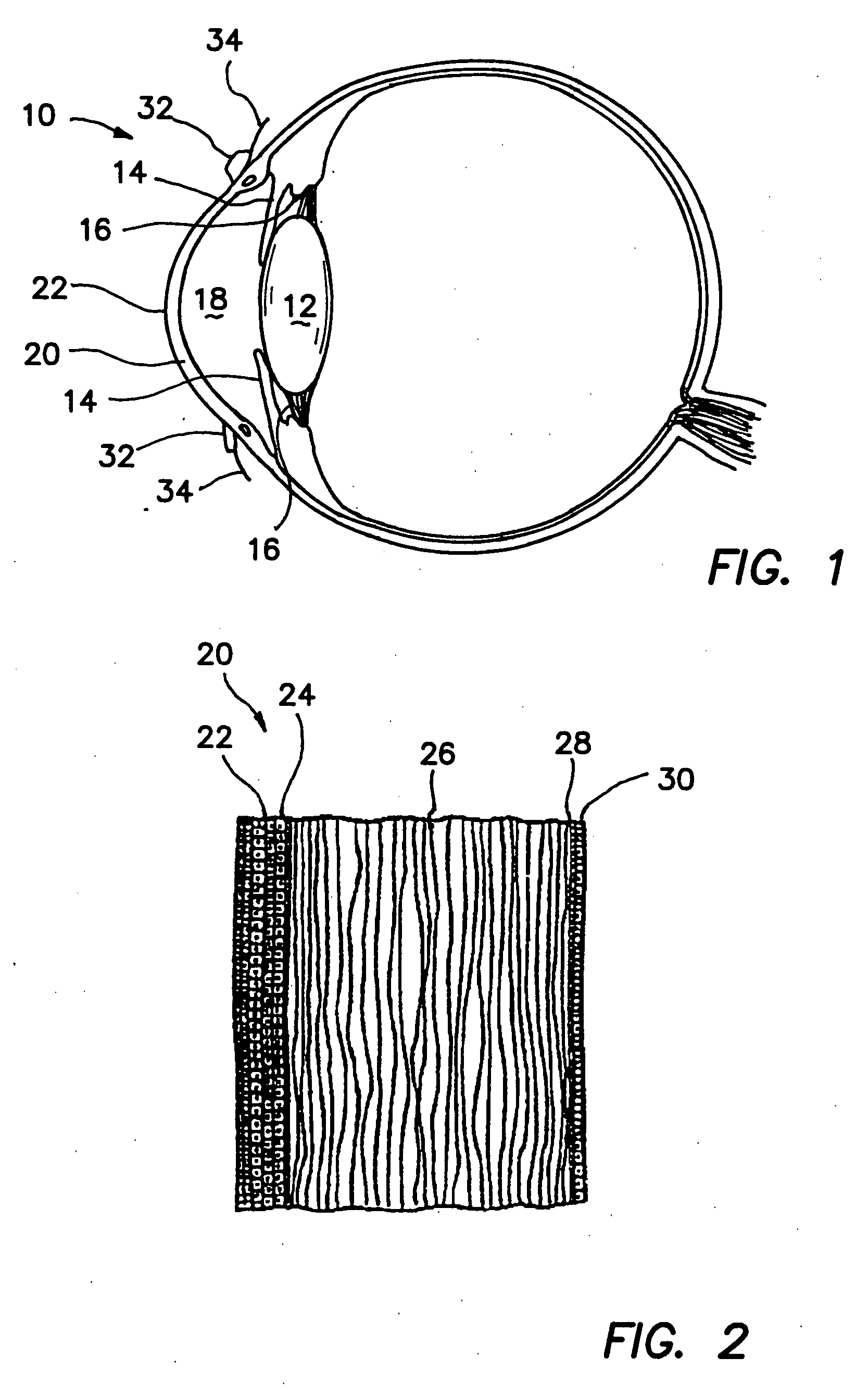
[0059] As illustrated in FIG. 1, a typical human eye 10 has a lens 12 and an iris 14. Posterior chamber 16 is located posterior to iris 14 and anterior chamber 18 is located anterior to iris 14. Eye 10 has a cornea 20 that consists of five layers, as discussed herein. One of the layers, corneal epithelium 22, lines the anterior exterior surface of cornea 20. Corneal epithelium 22 is a stratified squamous epithelium that extends laterally to the limbus 32. At limbus 32, corneal epithelium 22 becomes thicker and less regular to define the conjunctiva 34.

[0060]FIG. 2 illustrates a magnified view of the five layers of cornea 20. Typically, cornea 20 comprises corneal epithelium 22, Bowman's membrane 24, stroma 26, Descemet's membrane 28, and endothelium 30. Corneal epithelium 22 usually is about 5-6 cell layers thick (approximately 50 micrometers thick), and generally regenerates when the cornea is injured. Corneal epithelium 22 provides a relatively smooth refractive surface and helps...


PUM


Abstract

A corneal appliance that is placed over an eye has a lens body and epithelial cells secured over the lens body. The epithelial cells of the appliance may be derived from cultured cells, including stem cells, such as limbal stem cells, or epithelial cell lines, or may include at least a portion of the epithelium of the eye on which the appliance is placed. The corneal appliance may have a cellular attachment element between the lens body and the epithelial cells to facilitate attachment of the epithelial cells over the lens body. The corneal appliance is intended to be used on a deepithelialized eye, which may be an eye that has had the epithelium fully or partially removed. The corneal appliance may be used to improve vision. Methods of producing the corneal appliance and of improving vision are also disclosed.

Description

CROSS-REFERENCE TO RELATED APPLICATIONS [0001] This application claims the benefit of U.S. Provisional Application No. 60 / 464,590, filed Apr. 21, 2003, and U.S. Provisional Application No. 60 / 464,004, filed Apr. 18, 2003, and U.S. Provisional Application No. 60 / 410,837, filed Sep. 13, 2002, the disclosures of all of which are hereby incorporated by reference.BACKGROUND OF THE INVENTION [0002] 1. Field of the Invention [0003] The present invention relates to devices and methods of improving a patient's vision. In particular, the invention relates to improving vision of a patient by placing a corrective ocular device between an epithelium of the patient's eye and the stroma of the cornea of the patient's eye. The corrective ocular device may be a lens, including a corneal onlay. The corrective ocular device may have a preformed epithelial cell layer secured over the device when placed on an eye of a patient. The preformed epithelial cell layer may be synthesized in vitro or the prefor...

Claims


Application Information

Patent Timeline
IPC IPC(8): A61F2/14A61F9/00A61F9/013A61L27/38
CPCA61F2/145A61F9/0017A61F9/013A61L27/3604Y10S623/906A61L27/3813A61L27/3839A61L27/3895A61L2430/16A61L27/3641A61F2/14A61F2/1453
Inventor MARMO, J. CHRISTOPHERBACK, ARTHUR
Owner FORSIGHT LABS
Why Eureka
Try Eureka