Large-area imaging by stitching with array microscope

an array microscope and microscope technology, applied in the field of microscopy, can solve the problems of inconvenient operation, high cost, and inability to image large areas with high resolution, and achieve the effect of facilitating rapid and accurate operation

Inactive Publication Date: 2005-04-21
DMETRIX INC
View PDF11 Cites 38 Cited by
  • Summary
  • Abstract
  • Description
  • Claims
  • Application Information

AI Technical Summary

Benefits of technology

[0013] Thus, by applying the resulting correction-factor matrix to the data acquired by scanning the sample object in step-and-repeat fashion, the resulting checkerboard images are normalized to a uniform basis so that they can be concatenated or combined by stitching without further processing. As a result of this normalization process, the concatenation or stitching operation can be advantageously performed rapidly and accurately for the entire composite image simply by aligning pairs of adjacent images from the image checkerboards acquired during the scan. A single pair of images from each pair of checkerboards is sufficient because the remaining images are automatically aligned as well to produce a uniform result by virtue of their fixed spatial position within the checkerboard.

Problems solved by technology

Typical microscope objectives suffer from the inherent limitation of only being capable of imaging either a relatively large area with low resolution or, conversely, a small area with high resolution.
Therefore, imaging large areas with high resolution is problematic in conventional microscopy and this limitation has been particularly significant in the field of biological microscopy, where relatively large samples (in the order of 20 mm×50 mm, for example) need to be imaged with very high resolution.
Multi-element lenses with a large field of view and a high numerical aperture are available in the field of lithography, but their cost is prohibitive and their use is impractical for biological applications because of the bulk and weight associated with such lenses.
The absolute magnification in an array microscope is greater than one, which means that it is not possible to image the entire object surface at once even when it is equal to or smaller than the size of the array.
The patent does not provide any teaching regarding the way such multiple sets of checkerboard images may be combined to produce a high-quality high-resolution composite image.
In fact, while stitching techniques are well known and used routinely to successfully combine individual image tiles, the combination of checkerboard images presents novel and unique problems that cannot be solved simply by the application of known stitching techniques.
For example, physical differences in the structures of individual miniaturized objectives and tolerances in the precision with which the array of microscopes is assembled necessarily produce misalignments with respect to a common coordinate reference.
Moreover, optical aberrations and especially distortion and chromatic aberrations, as well as spectral response and gain / offset properties, are certain to vary from microscope to microscope, thereby producing a checkerboard of images of non-uniform quality and characteristics.
Therefore, the subsequent stitching by conventional means of multiple checkerboards of image tiles acquired during a scan cannot produce a high-resolution composite image that precisely and seamlessly represents the sample surface.
The combination of images acquired with different microscopes, though, could not be carried out meaningfully with conventional stitching techniques.
Therefore, the overall composite image could represent a meaningless assembly of incompatible image tiles that are incapable of producing an integrated result (like combining apples and oranges).
Thus, the prior art does not provide a practical approach to the very desirable objective of imaging a large area with an array microscope in sequential steps to produce checkerboards of images that can later be combined in a single operation simply by aligning any pair of adjacent image tiles.
Similarly, the prior art does not provide a solution to the same problem of image non-uniformity produced by an array microscope that is scanned linearly over a large area of the sample surface to produce image swaths that are later combined to form a composite image.

Method used


Image

  • Large-area imaging by stitching with array microscope
  • Large-area imaging by stitching with array microscope
  • Large-area imaging by stitching with array microscope

Examples


Embodiment Construction

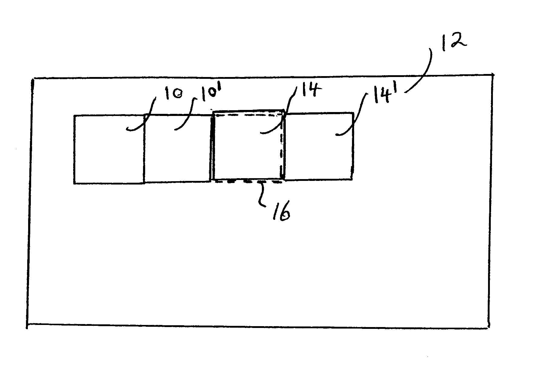
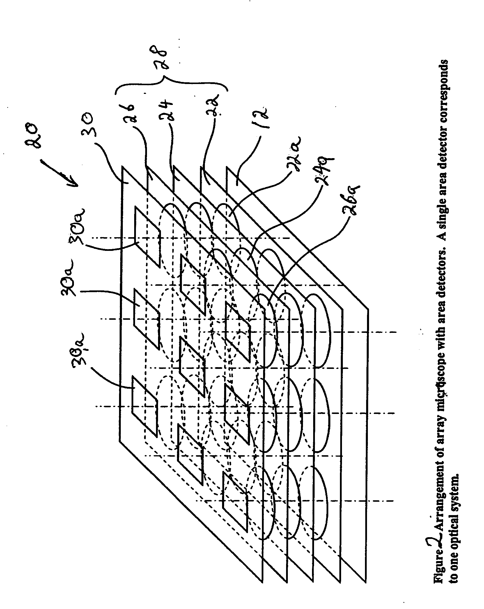
[0027] The invention was motivated by the realization that the images produced by step-and-repeat data acquisition using an array microscope cannot be combined directly to produce a uniform composite image because of the unavoidable data incompatibilities produced by discrepancies in the optical properties of the various miniaturized microscopes in the array. The heart of the invention lies in the idea of normalizing such optical properties to a common basis, so that functionally the array of microscopes performs, can be viewed, and can be treated as a single optical device of uniform characteristics. As a result, each set of multiple checkerboard images produced simultaneously at each scanning step can be viewed and treated as a single image that can be aligned and stitched in conventional manner with other sets in a single operation to produce the composite image of a large area.

[0028] As development of the invention progressed, it became apparent that the same advantages provide...


PUM


Abstract

An imaging apparatus consists of multiple miniaturized microscopes arranged into an array capable of simultaneously imaging a portion of an object. A step-and-repeat approach is followed to scan the object and generate multiple sets of checkerboard images. In order to improve the quality of the composite image produced by concatenation or stitching of the checkerboard images, the performance of each microscope is normalized to the same base reference for each relevant optical-system property. Correction factors are developed through calibration to equalize the spectral response measured at each detector; to similarly balance the gains and offsets of the detector / light-source combinations associated with the various objectives; to correct for geometric misalignments between microscopes; and to correct optical and chromatic aberrations in each objective.

Description

BACKGROUND OF THE INVENTION [0001] 1. Field of the Invention [0002] This invention is related in general to the field of microscopy. In particular, it relates to array microscopes and to a novel approach for acquiring multiple sets of image tiles of a large sample area using an array microscope and subsequently combining them to form a good-quality high-resolution composite image. [0003] 2. Description of the Related Art [0004] Typical microscope objectives suffer from the inherent limitation of only being capable of imaging either a relatively large area with low resolution or, conversely, a small area with high resolution. Therefore, imaging large areas with high resolution is problematic in conventional microscopy and this limitation has been particularly significant in the field of biological microscopy, where relatively large samples (in the order of 20 mm×50 mm, for example) need to be imaged with very high resolution. Multi-element lenses with a large field of view and a high...

Claims


Application Information

Patent Timeline
Patent Type & Authority Applications(United States)
IPC IPC(8): G02B21/36G06K9/36G06T3/40
CPCG02B21/365G06T3/4038G06K2009/2045G06K9/00134G02B21/367H04N1/3876G06V20/693G06V10/16
Inventor OLSZAK, ARTUR G.
Owner DMETRIX INC
Why Eureka
Try Eureka