

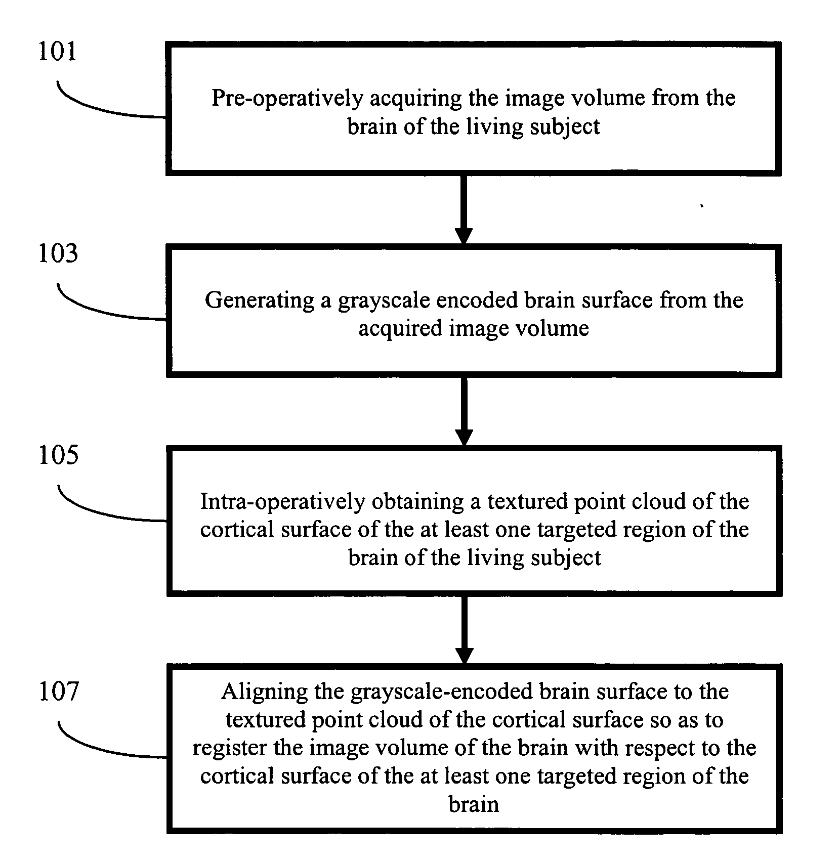
Apparatus and methods of cortical surface registration and deformation tracking for patient-to-image alignment in relation to image-guided surgery
a technology of image-guided surgery and apparatus, which is applied in the field of apparatus and methods of cortical surface registration and/or deformation tracking for patient-to-image alignment in relation to image-guided surgery, can solve the problems that systematic evaluation has not been performed to date, and achieve the effect of facilitating diagnostic or surgical procedures
- Summary
- Abstract
- Description
- Claims
- Application Information
AI Technical Summary
Benefits of technology
Problems solved by technology
Method used
Image


Examples
example 1
Phantom Experiment
[0082] To evaluate the accuracy and effectiveness of the SurfaceMI algorithm to register intermodality surfaces, a phantom experiment using a watermelon was conducted. Referring to FIGS. 5a-5c, in the experiment, Omnipaque (Amersham Health PLC, Buckinghamshire, the United Kingdom) soaked twine 510 was laid into the watermelon surface 520 to simulate the appearance of contrast-enhanced vasculature on the brain surface in CT, and / or MR imaging. Rigid fiducial markers 530, such as Acustar® (Z-Kat, Inc., Hollywood, Fla.), were implanted into the watermelon surface 520 for alignment of one image space to another one. The Acustar® fiducial markers 530 were filled with CT and / or MR visible contrast enhancement liquid. In addition, Acustar® divot caps 540 were placed at soaked twine (vessel) bifurcations for target localization. The phantom was imaged by a CT imager, such as Mx8000, (Philips Medical Systems, Bothell, Wash.), and scanned by a LRS, e.g., RealScan3D, and dig...
example 2
Clinical Trials
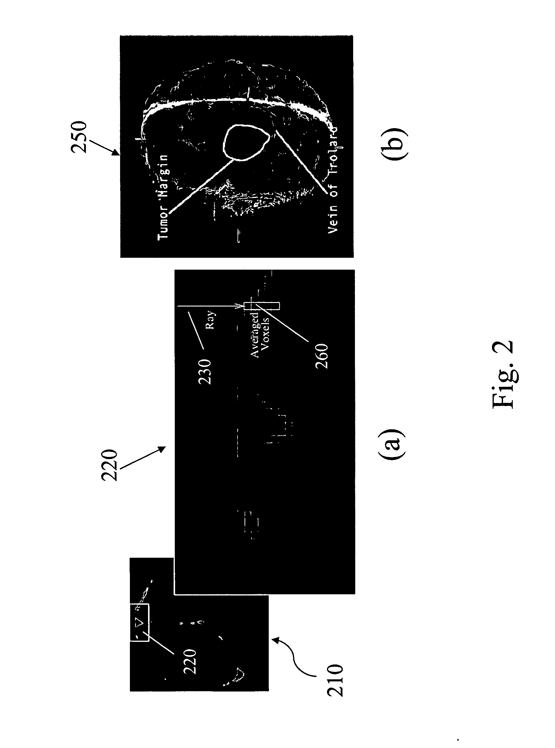
[0094] For clinical data acquisitions, the LRS approved by the VUIRB uses a Class I “eye-safe” laser and is mounted on a vibration damping monopod. Immediately after duratomy, the LRS is brought into the surgical FOV and the scanning extents (left and right scanning margins) are set to cover the width of the craniotomy. A laser stripe is then passed over the FOV and sampled 500 times between extents. Each sampling of the line produces 256 3D sample points, which results a practical point cloud density of approximately 100,000 points per scan. Immediately following the range scan, an RGB-bitmap (texture) of the scanning FOV is acquired and texture map coordinates are assigned to each 3D point via manufacturer calibration. Data acquisition by the LRS takes on the order of 15 to 30 seconds, with approximately 1.5 minutes of intra-operative time spent per scan (i.e. bring the LRS into the surgical FOV, calibrate extents and acquire, then remove from the FOV). Clinical da...
example 3
Tracking of Surface Deformations
[0106] Among other things, the present invention also provides a non-rigid registration capability for the tracking of brain surface deformations with serial range scans.
[0107] The data acquisition procedure for tracking brain surface deformations with serial range scans is described above. A laser range scanning device, for example, RealScan3D, was used to intra-operatively capture a 3D topography of the surgical FOV as well surface texture mapping to submillimeter accuracy, which describes the deforming nature of the brain during surgery. This scanner was mounted on a vibration-damped monopod that was brought into and out of the surgical FOV manually. After dural opening, the monopod and scanner were brought into the surgical FOV and the laser scanning extents (left and right margins) were calibrated to cover the width of the craniotomy. A laser stripe was then passed over the brain's surface and range data was collected using the principle of opt...
PUM
 Abstract
Description
Claims
Application Information