Compounds, methods of screening, and in vitro and in vivo uses involving anti-apoptotic genes and anti-apoptotic gene products

a gene and anti-apoptosis technology, applied in the field of cell physiology, can solve the problems of inappropriate or premature cell death or inappropriate cell proliferation, limited treatment options available, viral polypeptides that did not suppress apoptosis, etc., and achieve the effect of enhancing the stability, growth and/or productivity of cells

Inactive Publication Date: 2005-09-01
IMMUNOGEN INC
View PDF3 Cites 18 Cited by
  • Summary
  • Abstract
  • Description
  • Claims
  • Application Information

AI Technical Summary

Benefits of technology

[0055] Another embodiment of the present invention provides methods of enhancing the stability, growth, and / or productivity of cells by introducing into cells, by, for example, transfection or retrovirus infection, a polynucleotide encoding a polypeptide having anti-apoptotic activity, and expressing the polypeptide in the cells; wherein the cells under normal conditions do not express such a polypeptide. Examples of such cells are hybridomas, Chinese Hamster Ovary (CHO) cells, fibroblasts, lymphoid cells, haematopoietic cells, cells derived from the embryonic central nervous system, and cells derived from normal, dysplastic, or neoplastic tissue.
[0056] In particular, an embodiment of the present invention provides methods of enhancing the stability, growth, and / or productivity of cells by introducing into cells, by transfection or retrovirus infection, a polynucleotide encoding at least one HCMV polypeptide pUL36, pUL37S, pUL37M, or pUL37L, or encoding at least one fragment thereof, or encoding a homologue of such HCMV polypeptides or fragment thereof, and expressing the polypeptide in the cells; wherein the cells under normal conditions do not express such a polypeptide.

Problems solved by technology

A variety of diseases and degenerative disorders may involve aberrant or disregulated apoptosis, resulting in inappropriate or premature cell death or inappropriate cell proliferation (Barr, P. J., et al., 1994).
However, these viral polypeptides did not suppress apoptosis in other assays (see below).
Today, there are only very limited treatment options available for cytomegalovirus infections, and treatment is often associated with high toxicity and the generation of drug resistance (Hirsch, 1994; White and Fenner, 1994; Lalezari et al., 1997).
There are no available antiviral compounds that function to inhibit or diminish viral infection or to eliminate virally infected cells by regulating or modulating the anti-apoptotic activity of viral polypeptides and / or the expression of viral genes or polynucleotides encoding polypeptides having anti-apoptotic activity, and thereby inducing or restoring apoptosis in virally infected cells.

Method used


Image

  • Compounds, methods of screening, and in vitro and in vivo uses involving anti-apoptotic genes and anti-apoptotic gene products
  • Compounds, methods of screening, and in vitro and in vivo uses involving anti-apoptotic genes and anti-apoptotic gene products
  • Compounds, methods of screening, and in vitro and in vivo uses involving anti-apoptotic genes and anti-apoptotic gene products

Examples


experimental examples

[0343] The invention will now be illustrated by reference to non-limiting examples. Unless otherwise stated, all percents, ratios, parts, etc. are by weight.

example 1

Induction and Detection of Apoptotic Activity.

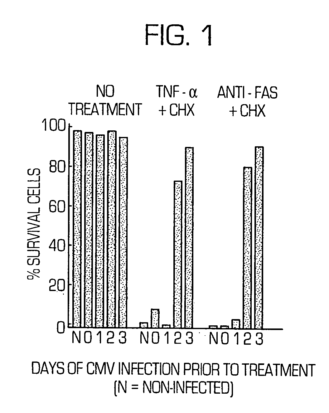
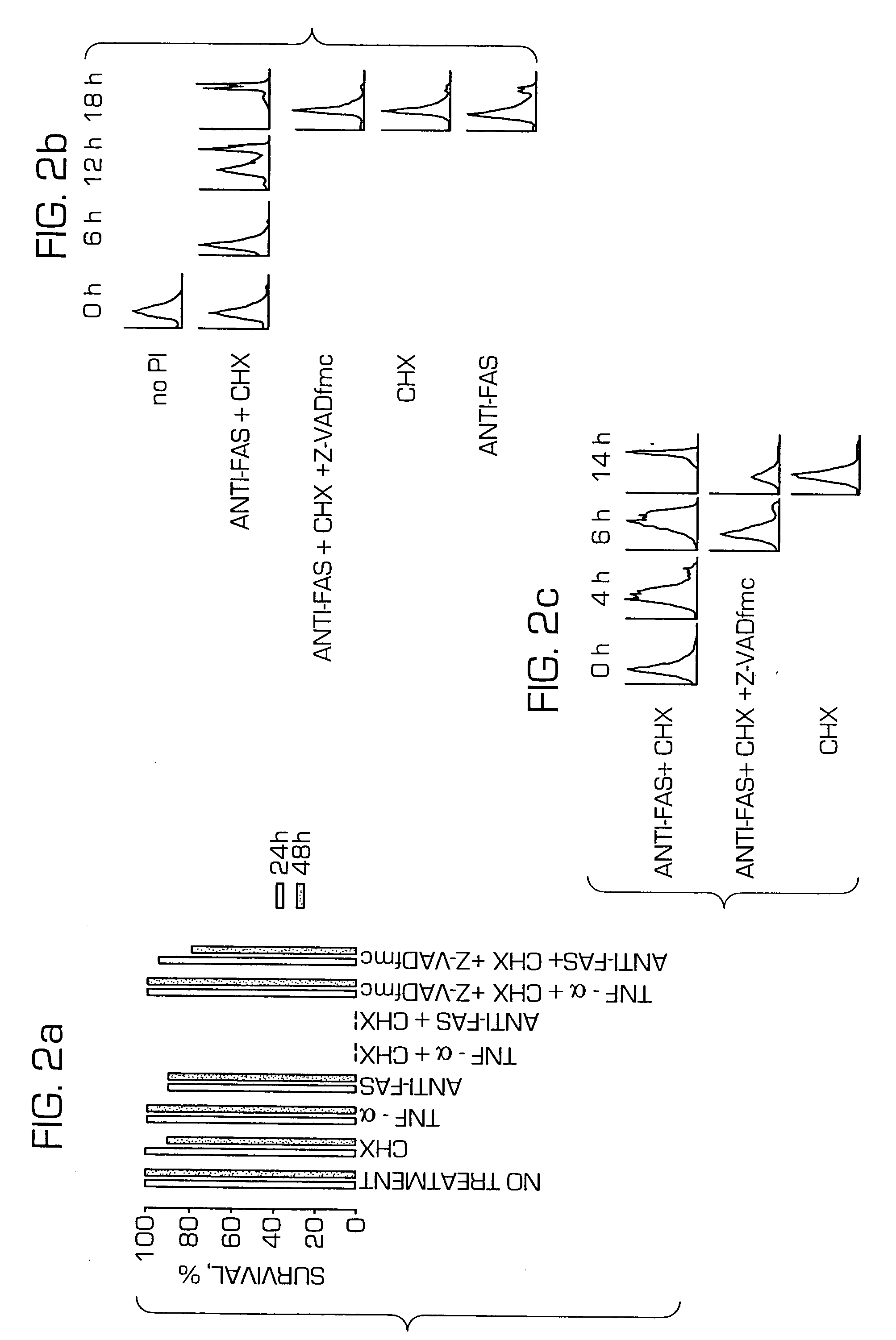
[0344] Tumor necrosis factor receptor 1 (TNF-R1)-mediated apoptosis was induced by exposure of HeLa and MRC-5 cells to tumor necrosis factor-α (TNF-α) (Sigma, 10-60 ng / mL)+CHX (10-30 μg / mL). Fas-mediated apoptosis was induced by exposure of cells to an anti-Fas antibody (Coulter, 7C11 antibody at 0.2-4 μg / mL)+CHX (10-30 μg / mL). The degree of cell death was determined by the visual scoring of surviving cells, in representative fields, under a phase microscope. A similar degree of cell death occurred in fibroblasts when examined 5 to 28 h after the start of exposure, or a similar degree of cell death occurred in HeLa cells when examined 9-28 h after the start of exposure. Further, a similar degree of cell death for a given reagent was produced when any concentration within the range indicated above was used.

[0345] The time-course of apoptosis was also analyzed by examining the expression of phosphatidylserine on the cell surface, as det...

example 2

Detection of Intracellular Polypeptides.

[0349] Cells were trypsinized, washed in Tris buffered saline, resuspended in a lysis buffer (150 mM NaCl, 5 mM EDTA, 50 mM Tris.HCl pH 8.0, 1% Triton X-100), freshly supplemented with phenylmethylsulfonyl fluoride (0.1 mM), leupeptin (1 μg / mL) and pepstatin (1 μg / mL), incubated on ice for 30 minutes, and centrifuged at 16,000 g for 15 minutes, at 4° C. Cytochrome c and procaspase 9 were detected in S-100 fractions of cells (Li et al., 1997, which is incorporated herein by reference).

[0350] The intracellular polypeptides were detected by Western blot analysis. Samples containing protein were electrophoretically resolved under reduced conditions, on Laemmli SDS-PAGE (Novex), after equal amounts of protein were loaded per well of the gel, detected using a standard Western blot protocol and a luminol-based ECL Western Blotting system (Amersham). The following antibodies were used in the Western blot analysis: anti-human Bid C-20 (Santa Cruz Bi...


PUM

PropertyMeasurementUnit
temperatureaaaaaaaaaa
pHaaaaaaaaaa

Abstract

Novel polypeptides having anti-apoptotic activity, and methods of screening for such novel polypeptides having anti-apoptotic activity, and polynucleotides encoding such polypeptides; Compounds that regulate or modulate apoptosis and / or anti-apoptotic activity, such as compounds having anti-apoptotic activity, and such as compounds that induce, restore, or modulate apoptosis and / or inhibit, diminish, or modulate anti-apoptotic activity, methods of screening for such compounds, and methods of using such compounds in the therapeutic treatment of diseases; Methods of treating eukaryotic cells with compounds that regulate or modulate apoptosis and / or anti-apoptotic activity; Methods of enhancing the stability, growth, and / or productivity of eukaryotic cells; Pharmaceutical compositions that regulate or modulate apoptosis and / or anti-apoptotic activity.

Description

[0001] The instant application is a Continuation-In-Part Application of U.S. application Ser. No. 09 / 301,121, filed Apr. 28, 1999, which is a Continuation-In-Part Application of U.S. application Ser. No. 08 / 080,265, filed May 18, 1998, each of which is incorporated herein by reference.FIELD OF THE INVENTION [0002] The present invention relates generally to the field of cell physiology, and more particularly, to apoptosis. More specifically, the present invention relates to methods for detecting the anti-apoptotic activity and function of viral polypeptides and their interactions with cellular polypeptides. The present invention further relates to viral polypeptides having anti-apoptotic activity and also the mechanism by which such viral polypeptides function to regulate or modulate apoptosis or cell death. Moreover, the present invention relates to compounds that regulate or modulate the anti-apoptotic activity of viral polypeptides, and expression of polynucleotides encoding such ...

Claims


Application Information

Patent Timeline
Patent Type & Authority Applications(United States)
IPC IPC(8): A61B17/80A61B17/86A61F2/00A61F2/02A61F2/30A61F2/38A61F2/46A61K31/7088A61K38/00A61K39/395A61K48/00A61P9/10A61P31/00A61P31/12A61P35/00A61P37/00A61P37/06A61P43/00C07H21/04C07K14/045C07K16/08C12N15/09C12P21/08C12Q1/02C12Q1/68C12Q1/70G01N33/15G01N33/50G01N33/68
CPCA61K38/00A61K48/00C07K14/005C12N2710/16122G01N2510/00G01N33/5011G01N33/5017G01N33/502G01N33/5035G01N33/5008A61P9/10A61P31/00A61P31/12A61P35/00A61P37/00A61P37/06A61P43/00
Inventor GOLDMAKHER, VIKTORSKALETSKAYA, ANNABARTLE, LAURA
Owner IMMUNOGEN INC
Why Eureka
Try Eureka