Method and system for displaying regions of pathological interest

Inactive Publication Date: 2005-10-27
GE MEDICAL SYST INFORMATION TECH
View PDF15 Cites 32 Cited by
  • Summary
  • Abstract
  • Description
  • Claims
  • Application Information

AI Technical Summary

Problems solved by technology

Further, a high percentage of CAD-detect

Method used


Image

  • Method and system for displaying regions of pathological interest
  • Method and system for displaying regions of pathological interest
  • Method and system for displaying regions of pathological interest

Examples


Embodiment Construction

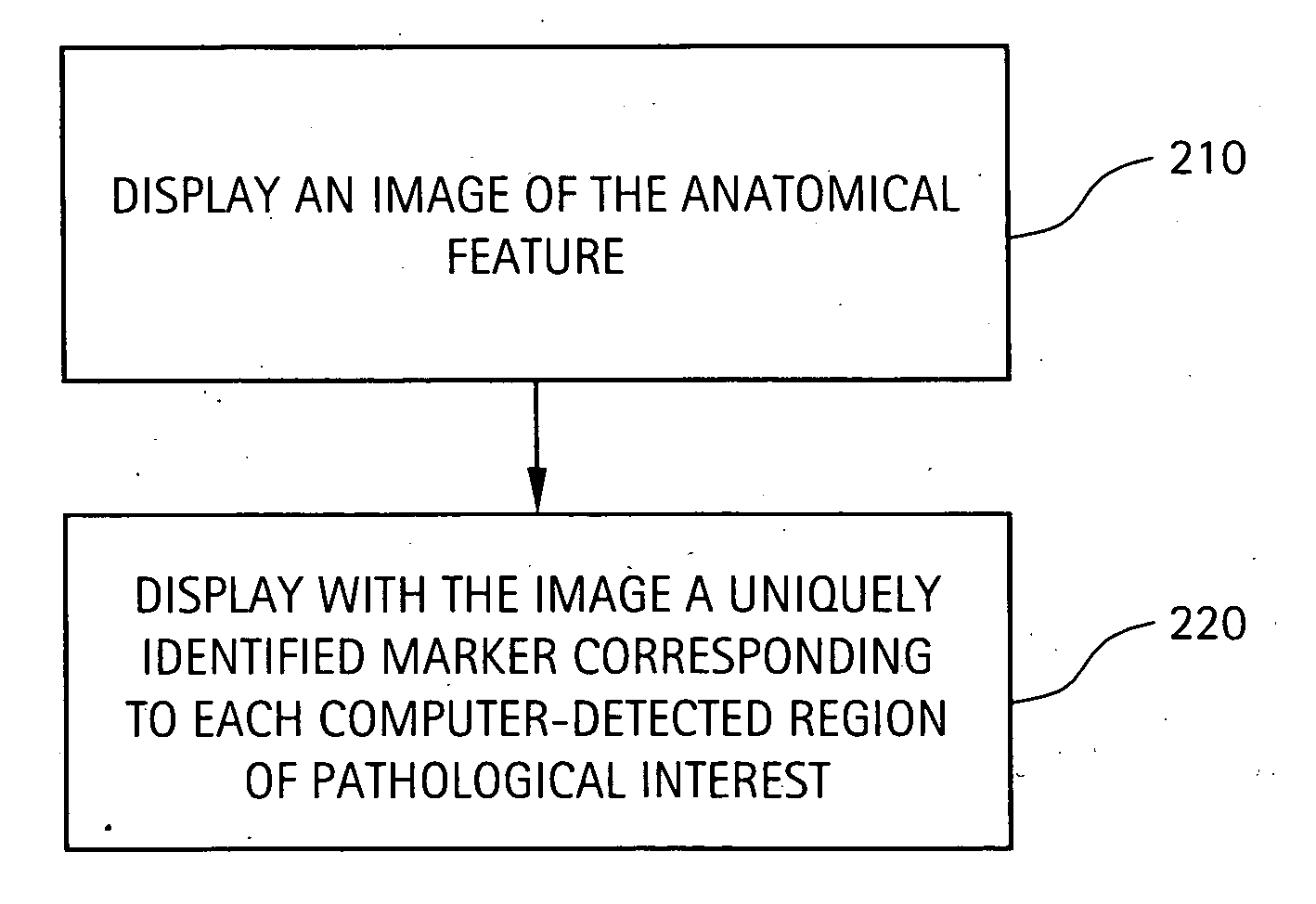
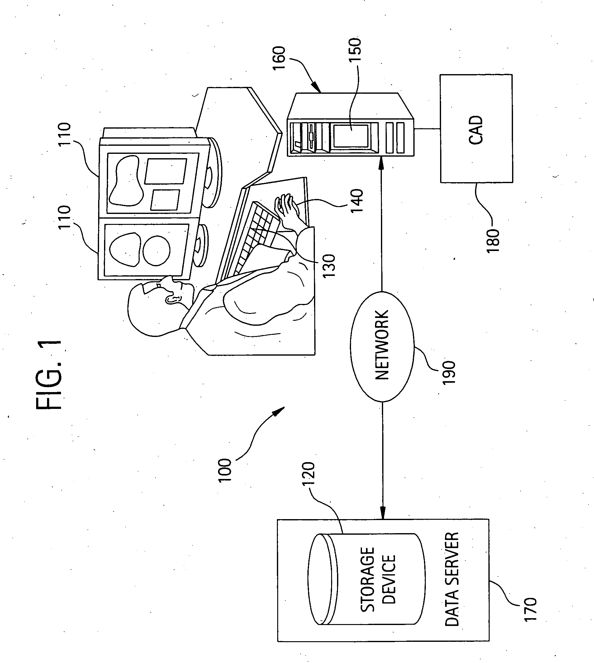
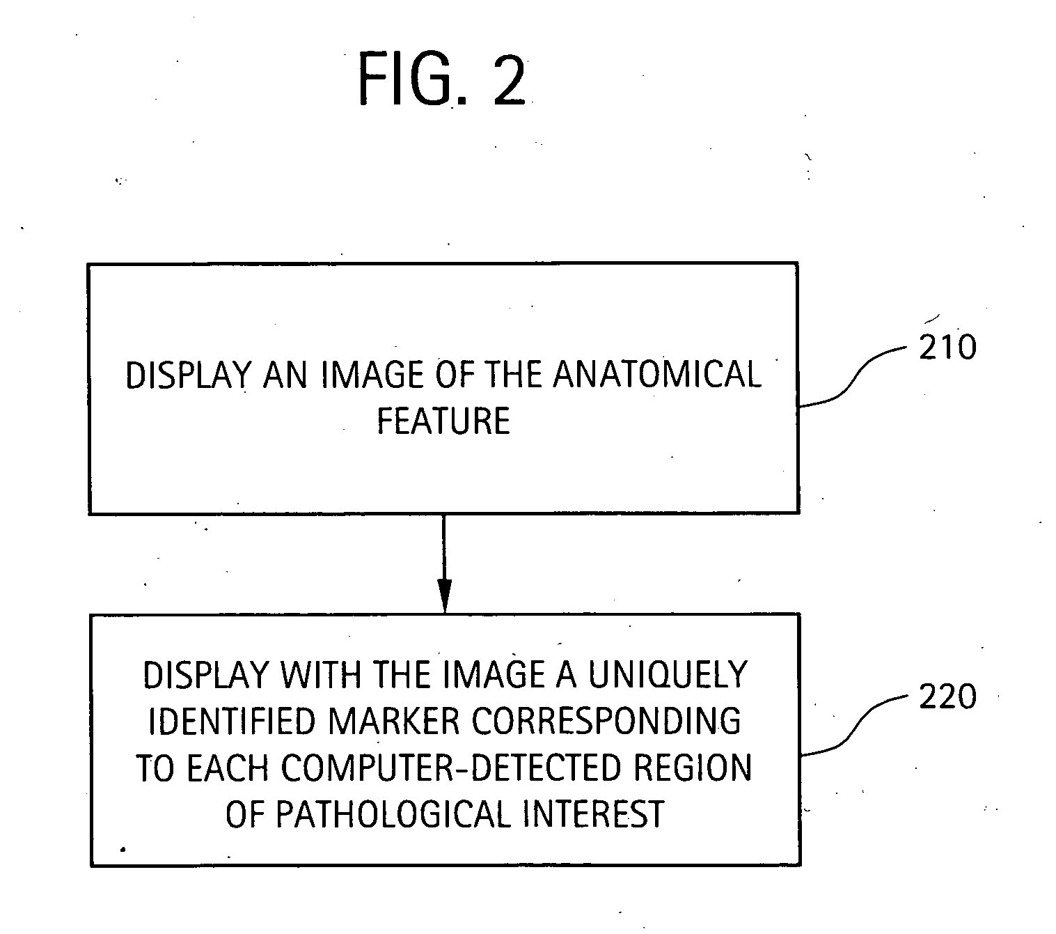
[0012] Turning now to the FIGURES which illustrate exemplary embodiments, a method and system are shown which may be used to display a number of computer-detected regions of pathological interest using interactive CAD markers. In one embodiment, a layer of uniquely identified CAD markers corresponding to computer-detected regions of pathological interest of an anatomical feature may be simultaneously displayed with an image of the anatomical feature. Each individual CAD marker may be configured to incorporate viewable classification data entered by a user and may further be configured to visually indicate the probability of cancer determined by a CAD algorithm. Each individual CAD marker and the information associated therewith may also be saved with the image such that it may be incorporated into, for example, a structured reporting system. These features allow for smoother workflow between, for example, radiologists and physicians, and improved ease of differentiation and prioriti...


PUM


Abstract

A method and system which may be used to display a number of computer-detected regions of pathological interest using interactive CAD markers are presented. In one embodiment, a layer of uniquely identified CAD markers corresponding to computer-detected regions of pathological interest of an anatomical feature may be simultaneously displayed with an image of the anatomical feature. Each individual CAD marker may be configured to incorporate viewable classification data entered by a user and may further be configured to visually indicate the probability of cancer determined by the CAD algorithm. Each individual CAD marker and the information associated therewith may also be saved with the image such that it may be incorporated into, for example, a structured reporting system.

Description

BACKGROUND OF THE INVENTION [0001] The present description relates generally to methods and systems for viewing medical images. In particular, the present description relates to a method and system for displaying identified regions of pathological interest using interactive CAD markers which contain unique visual identification data. [0002] Computer Aided Detection (CAD) technology is presently used in conjunction with various types of early detection cancer screening technologies, such as digital mammography, to assist radiologists and physicians in identifying suspicious lesions and other potential regions of pathological interest. CAD technology typically employs a computer-implemented algorithm to analyze an image of a particular anatomical feature, such as a breast or lung, and identify regions on the image of characteristics commonly associated with cancerous pathology. The CAD algorithm may also determine the probability of cancer for each detected region of the anatomical fe...

Claims


Application Information

Patent Timeline
IPC IPC(8): G06F9/00G16H30/40G16Z99/00
CPCG06F19/321G06K2209/05G06K9/033G06F19/345G16H50/20G16H30/40G16Z99/00G06V10/987G06V2201/03
Inventor MORITA, MARK M.FORS, STEVEN L.RAI, KHAL A.THOMAS, CARSON H.
Owner GE MEDICAL SYST INFORMATION TECH
Why Eureka
Try Eureka