Method to identify arterial and venous vessels

a technology of arterial and venous vessels and identifying methods, applied in the field of medical imaging, can solve the problem that the art lacks methods to ease the task of identifying the anatomical labels of vessels

Inactive Publication Date: 2005-11-17
THE BOARD OF TRUSTEES OF THE LELAND STANFORD JUNIOR UNIV
View PDF8 Cites 23 Cited by
  • Summary
  • Abstract
  • Description
  • Claims
  • Application Information

AI Technical Summary

Problems solved by technology

Currently the art lacks methods to ease the tas

Method used


Image

  • Method to identify arterial and venous vessels
  • Method to identify arterial and venous vessels
  • Method to identify arterial and venous vessels

Examples


Embodiment Construction

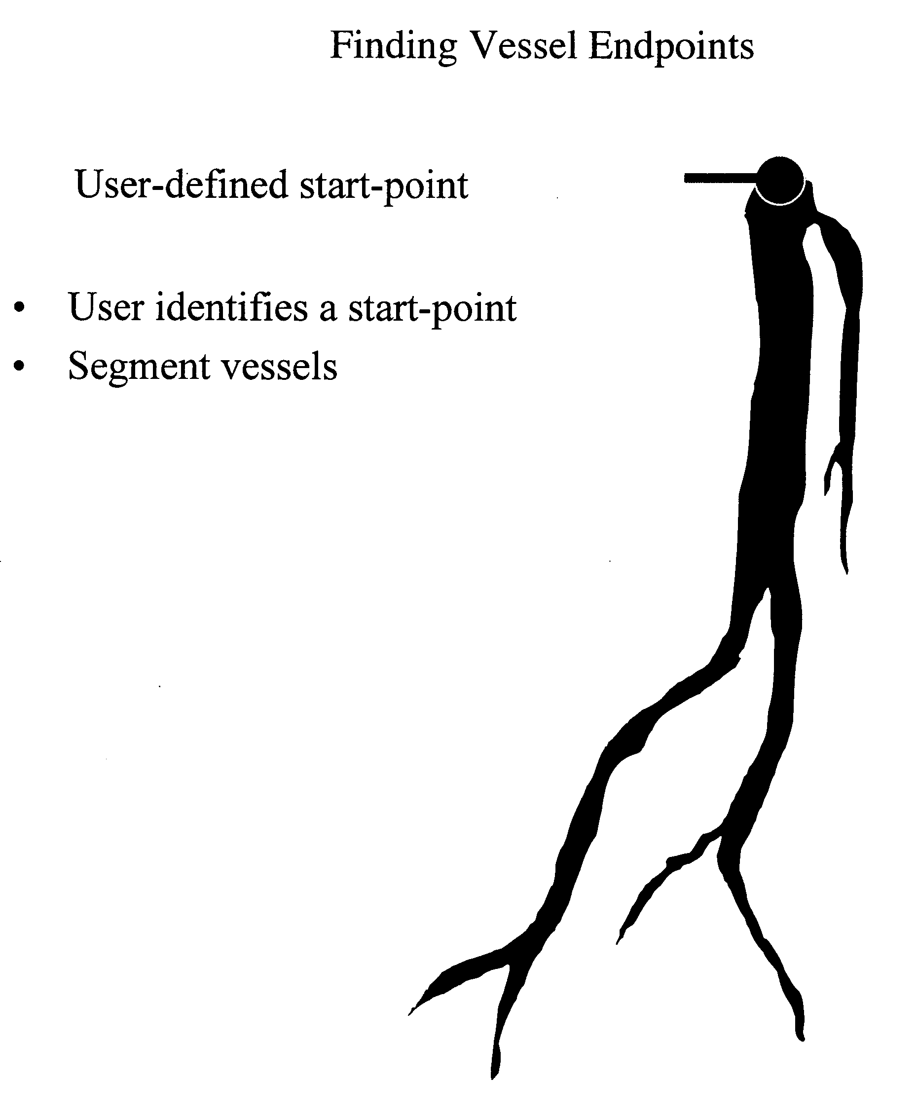
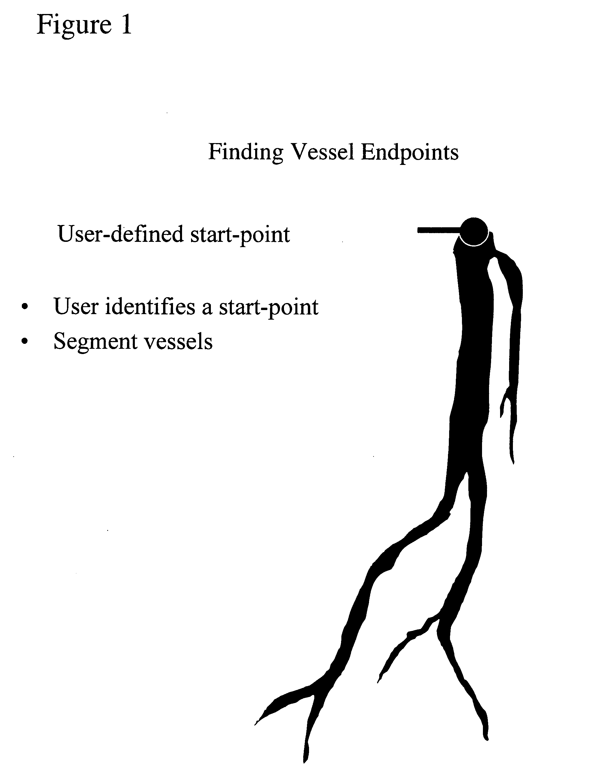
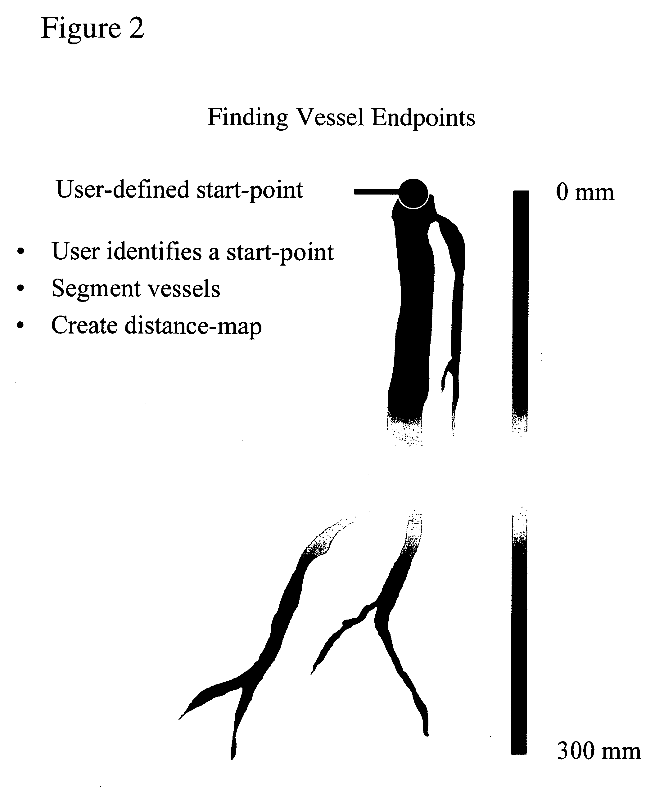
[0016] We have developed a method for automatically identifying the location and course of the vascular tree, given only one user-defined point in the root or parent vessel for the vascular tree. Our method also then uses the relatively invariant parameters (vessel cross sectional profile and vessel cross sectional area profile discontinuities, branching patterns, branch directions and laterality) of the human vascular tree to apply appropriate anatomic labels to the branches of the vascular tree.

[0017] The methods uses one (manually or automically) entered point in a vessel, e.g. the aorta, and patient orientation from the image headers to obtain position information for anatomic labeling. The method then creates a segmentation of the vessel tree. This is done by using an adaptive threshold and the startpoint as the seed point. The segmentation thus obtained represents the vascular tree in its entirety. A standard distancemap is then calculated. This distancemap enumerates the dis...


PUM


Abstract

A method for identifying a arteries and veins in a medical image is provided. A start point and endpoints of branches of a segmented tubular tree are identified. Distance maps for each of the endpoints relative to the startpoint are created. Then voxels in between the furthest of the endpoints and the startpoint are identified. This last step is iterated for the subsequent furthest of the endpoints. For each set of identified voxels parameters are identified. Examples of such parameters are cross sectional areas of the branches. The parameters for at least one each set of identified voxels are then used to anatomically label branches the segmented tubular tree, optionally with position information obtained from the image.

Description

CROSS-REFERENCE TO RELATED APPLICATIONS [0001] This application is cross-referenced to and claims priority from U.S. Provisional Application 60 / 526,560 filed Dec. 2, 2003. All these applications are hereby incorporated by reference. STATEMENT REGARDING FEDERALLY SPONSORED RESEARCH OR DEVELOPMENT [0002] The present invention was supported in part by grant numbers 5R01HL58915 and 1R01HL67194 both from the National Institutes of Health (NIH). The U.S. Government has certain rights in the invention.FIELD OF THE INVENTION [0003] The present invention relates generally to medical imaging. More particularly, the present invention relates to a method for identifying vessels. BACKGROUND [0004] Vessel structures in the body are routinely evaluated for vascular disease using imaging modalities such as Computed Tomographic Angiography (CTA) or Magnetic Resonance Angiography. The images thus obtained constitute a volumetric dataset which contains the vascular tree of the human body. The exact lo...

Claims


Application Information

Patent Timeline
IPC IPC(8): A61B5/05G06T7/00
CPCG06T2207/30101G06T7/0012
Inventor RAMAN, BHARGAVRAMAN, RAGHAVNAPEL, SANDY A.RUBIN, GEOFFREY D.
Owner THE BOARD OF TRUSTEES OF THE LELAND STANFORD JUNIOR UNIV
Why Eureka
Try Eureka