Clinical application of electrical impedance tomography to characterize tissue

Inactive Publication Date: 2006-01-05
KSN ENERGIES
View PDF8 Cites 53 Cited by
  • Summary
  • Abstract
  • Description
  • Claims
  • Application Information

AI Technical Summary

Benefits of technology

[0009] EIT clinical applications generally involve electrodes employed outside the body, electrically attached to the skin. EIT would be improved, however, through

Problems solved by technology

For example, systems such as x-rays and CAT scans expose the body to potentially harmful radiation.
Scanning with MRI may be da

Method used


Image

  • Clinical application of electrical impedance tomography to characterize tissue
  • Clinical application of electrical impedance tomography to characterize tissue
  • Clinical application of electrical impedance tomography to characterize tissue

Examples


Embodiment Construction

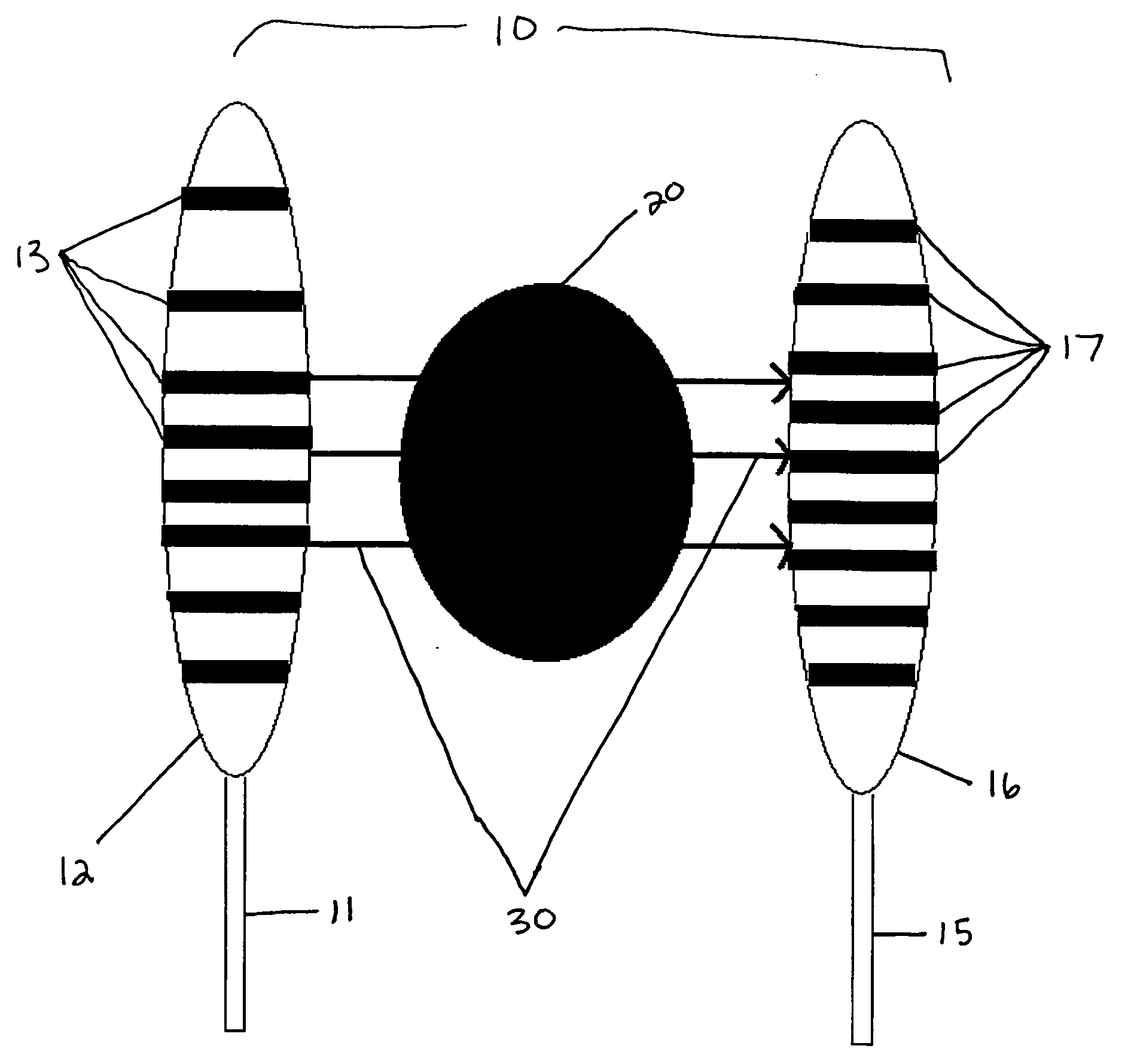
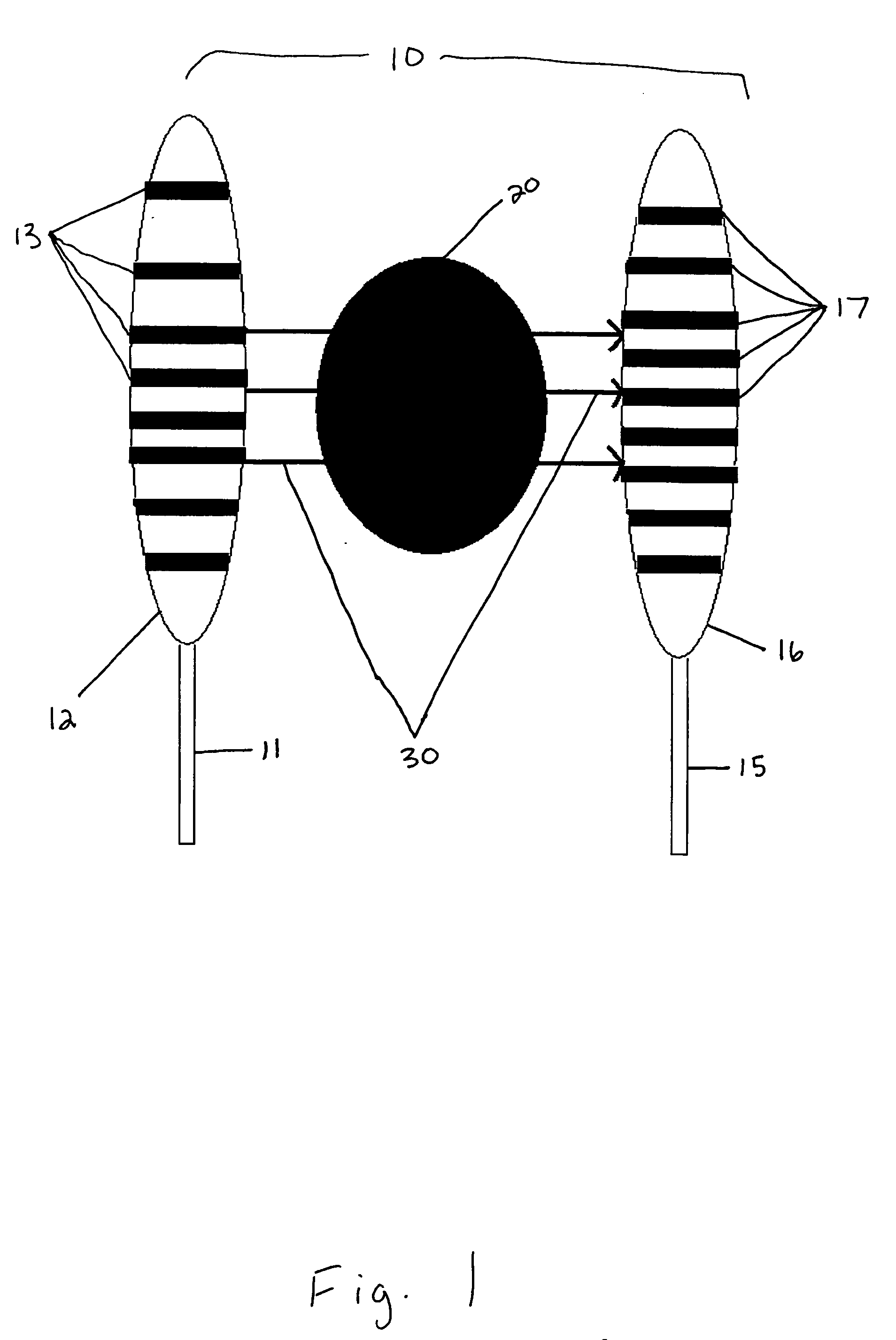
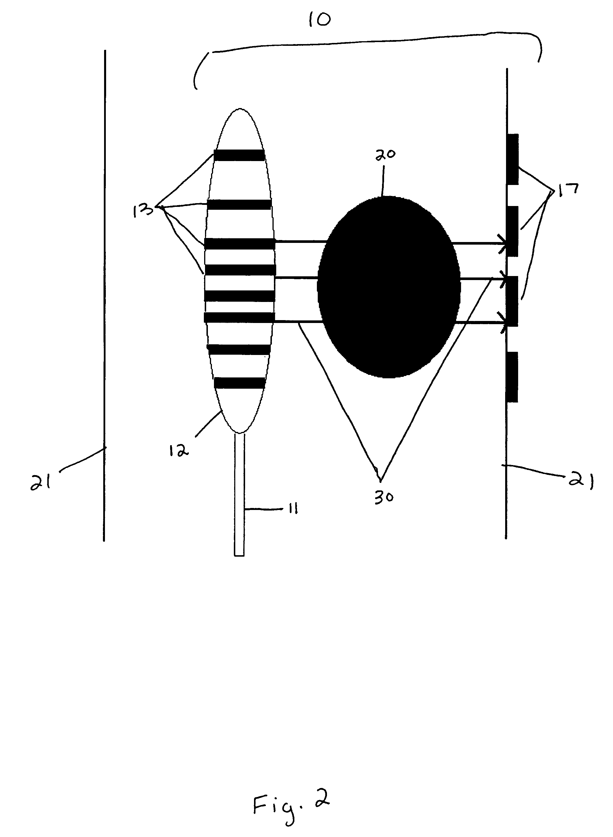
[0019] The present invention is a system and method for using electrical impedance tomography to characterize tissue in the human body. Any such system requires at least two sets of electrodes, one of current injection electrodes and one of current return electrodes. Voltages and currents may be applied to the electrode arrays, which creates a current from one to the other that runs through the intervening tissue. The system permits a measurement of the resistivity of the intervening tissue, which measurements are then used to create an image of the tissue which can be used to diagnose and / or treat disease or other conditions.

[0020] In the system and method of the current invention, one or both of the sets of electrodes are located inside the body during operation. For example, in one embodiment, the current injection electrode array is attached to the exterior of an expandable balloon. The expandable balloon is removably attached to the end of a flexible tube, such as a catheter, ...


PUM


Abstract

The present invention is a system for use in creating images of portions of human tissue inside the body by electrical impedance tomography. The system may comprise at least one flexible tube for insertion in the body in proximity to a targeted portion of human tissue in the body, an inflatable balloon removably attached to the distal end of the flexible tube, a first array of electrodes attached to the surface of the inflatable balloon for at least one of injecting current into the targeted portion of human tissue and receiving current that was injected into the targeted portion of human tissue, and a second array of electrodes for at least one of injecting current into the targeted portion of human tissue to the first array of electrodes and for receiving current from the first array of electrodes.

Description

CROSS-REFERENCE TO RELATED APPLICATION(S) [0001] This application claims priority from U.S. provisional patent application No. 60 / 582,720, which was filed on Jun. 24, 2004, and which is incorporated herein by reference in its entirety.FIELD OF THE INVENTION [0002] The present invention generally relates to the use of electrical impedance tomography (“EIT”) to characterize tissue in the human body. More particularly, this invention relates to systems for using EIT inside the human body to create high quality images of tissue to assist in the diagnosis and treatment of disease. BACKGROUND OF THE INVENTION [0003] There is a widespread need for high-quality images of human tissue in connection with diagnosing and treating diseases and other conditions. To name just a few examples, such imagery is invaluable for locating and treating tumors, diagnosing and locating pulmonary emboli, diagnosing and treating heart disease, and monitoring blood volume and blood flow. [0004] Traditional syst...

Claims


Application Information

Patent Timeline
IPC IPC(8): A61B5/05A61B5/053
CPCA61B5/0536A61B5/0538A61B5/4381A61B2562/046A61B2562/0209A61B2562/043A61B5/6853A61B2562/0215
Inventor KASEVICH, RAYMOND S.
Owner KSN ENERGIES
Why Eureka
Try Eureka