Biological tissue closure device and method

a technology of biological tissue and closure device, which is applied in the field of closing openings, can solve the problems of total occlusion of blood vessels, time-consuming methods, and discomfort of procedures for patients

Inactive Publication Date: 2006-01-12
ARSTASIS
View PDF69 Cites 86 Cited by
  • Summary
  • Abstract
  • Description
  • Claims
  • Application Information

AI Technical Summary

Problems solved by technology

This method is time-consuming, frequently requiring one-half hour or more of compression before hemostasis.
This procedure is uncomfortable for the patient and frequently requires administering analgesics.
Excessive pressure can also present the risk of total occlusion of the blood vessel, resulting in ischemia and / or thrombosis.
During this time bleeding from the vascular access wound can restart, potentially resulting in major complications.
This method generally presents difficulty locating the interface of the overlying tissue and the adventitial surface of the blood vessel.
Implanting the fastener too far from the desired location can result in failure to provide hemostasis.
If, however, the fastener intrudes into the vascular lumen, thrombus can form on the fastener.
Thrombus can embolize downstream and / or block normal blood flow at the thrombus site.
Implanted fasteners can also cause infection and auto-immune reactions / rejections of the implant.
Due to the lack of definite aiming of the acoustic transducer at the arteriotomy, the acoustic energy from the transducer can fail to seal the target arteriotomy, and / or can unintentionally effect surrounding tissue.
In addition, the arteriotomy is in the approximate shape of a cylinder, increasing the possibility that walls of the arteriotomy will be too far apart to seal together during energy application.

Method used


Image

  • Biological tissue closure device and method
  • Biological tissue closure device and method
  • Biological tissue closure device and method

Examples


Embodiment Construction

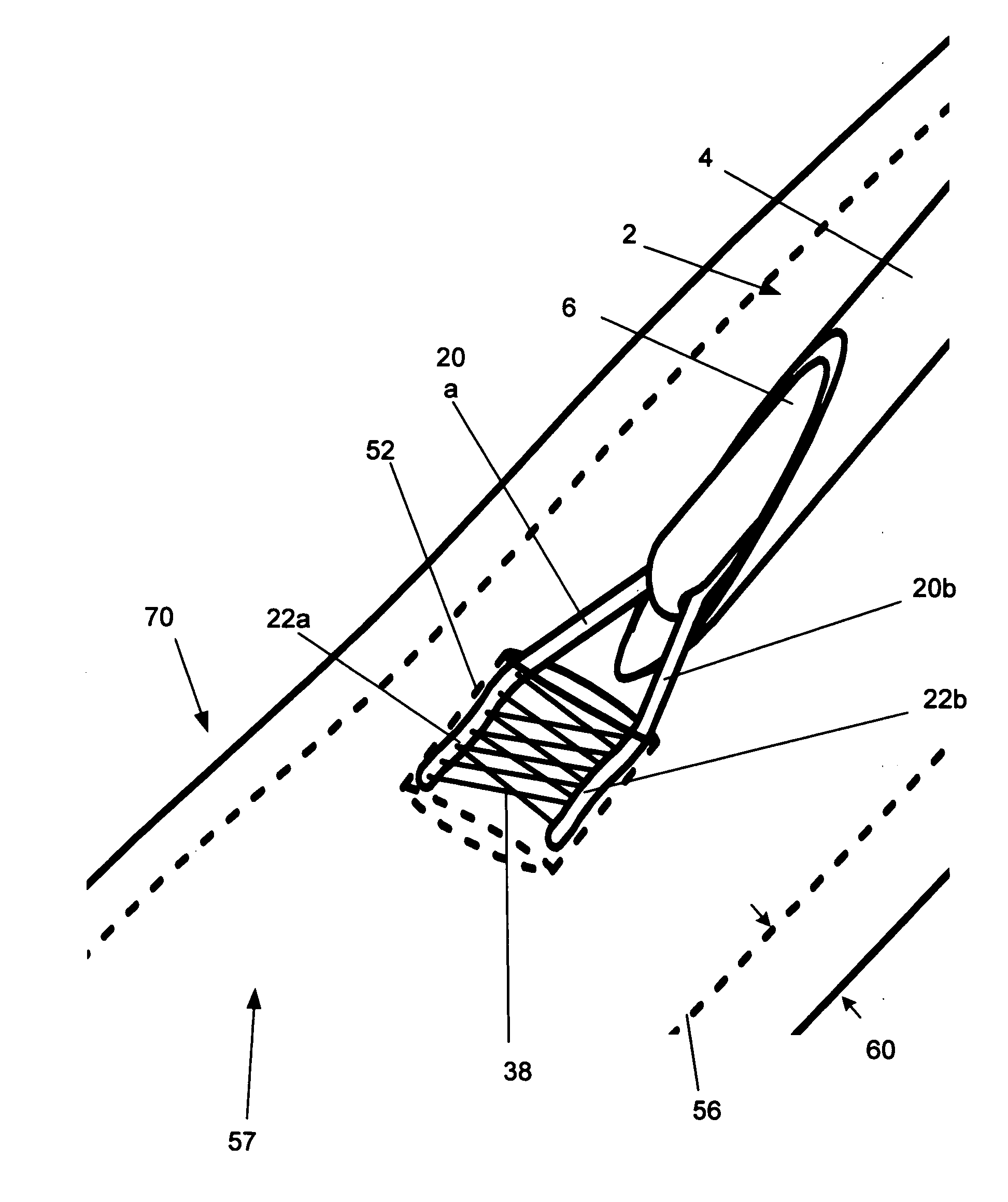
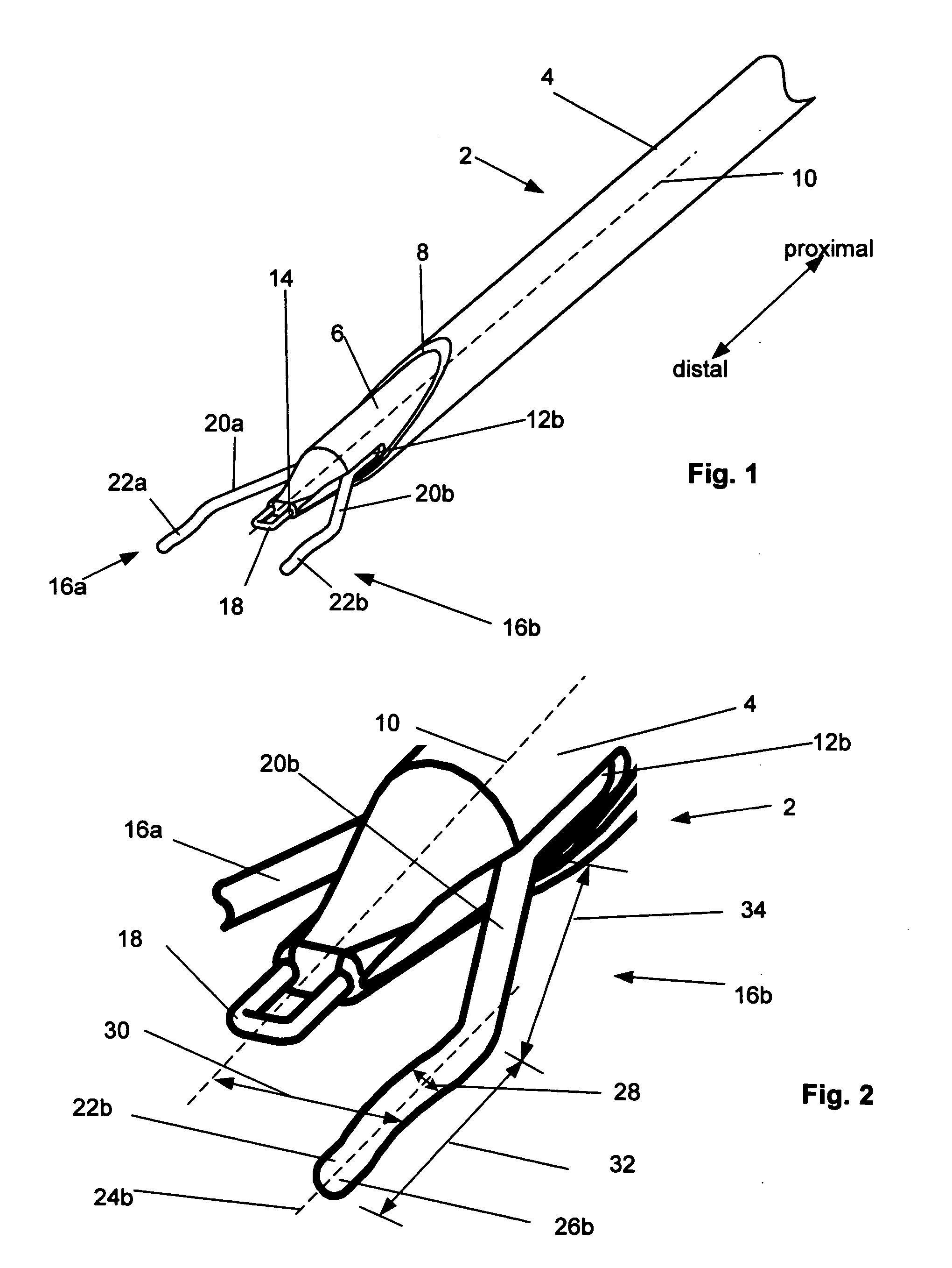
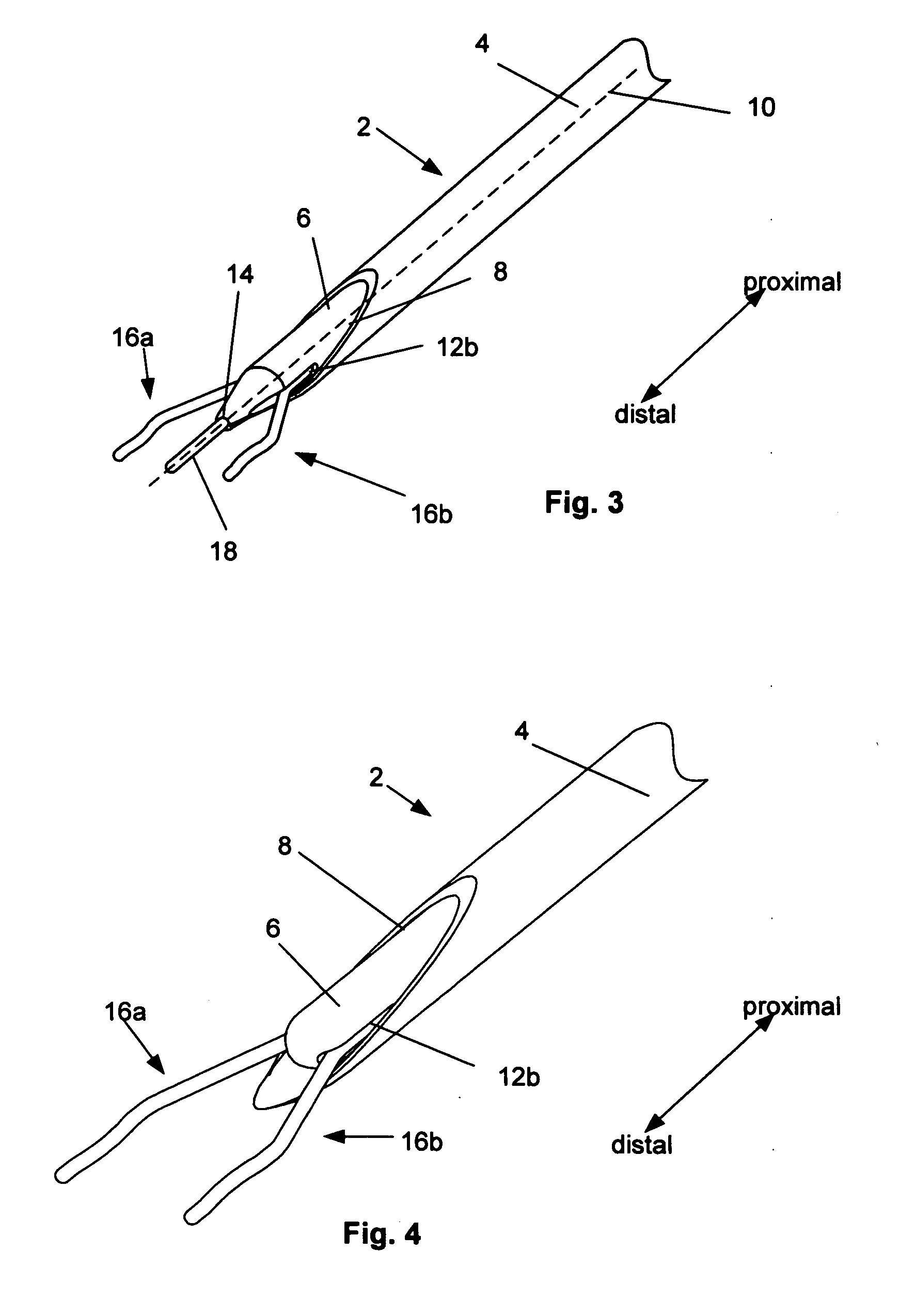
[0033]FIG. 1 illustrates in an extended (i.e., expanded) configuration, a closure device 2 for biological tissue closure, for example to create hemostasis across an arteriotomy. FIG. 2 illustrates a close-up of the distal end of the closure device of FIG. 1.

[0034] The closure device 2 can have a delivery guide 4. The delivery guide 4 can be a tubular member, such as a catheter or sheath on the outer radial side of the closure device 2. The delivery guide 4 can be hollow. In one configuration, the delivery guide 4 can be on the proximal end of the closure device 2. In another configuration, the delivery guide 4 can be the entire length of the closure device 2. The delivery guide 4 can have a low-friction inner surface. The delivery guide 4 can be configured to receive an inner member 6. The delivery guide 4 can have a distal port 8 at the distal end of the delivery guide 4.

[0035] The delivery guide 4 can have a proximally-located handle (not shown). The handle can facilitate manipu...


PUM


Abstract

Devices and methods for biological tissue closure are disclosed. Arteriotomy closure and hemostasis devices and methods are disclosed. A device that can provide a lateral tension across an opening in the tissue and apply energy to seal the tissue is disclosed. Methods for using the device are also disclosed.

Description

BACKGROUND OF THE INVENTION [0001] 1. Field of the Invention [0002] The present invention relates to the field of closing openings in biological tissue and methods of performing the same. [0003] 2. Description of the Related Art [0004] A number of diagnostic and interventional vascular procedures are now performed translumenally, where a catheter is introduced to the vascular system at a convenient access location—such as the femoral, brachial, or subclavian arteries—and guided through the vascular system to a target location to perform therapy or diagnosis. When vascular access is no longer required, the catheter and other vascular access devices must be removed from the vascular entrance and bleeding at the puncture site must be stopped. [0005] One common approach for providing hemostasis is to apply external force near and upstream from the puncture site, typically by manual compression. This method is time-consuming, frequently requiring one-half hour or more of compression befo...

Claims


Application Information

Patent Timeline
Patent Type & Authority Applications(United States)
IPC IPC(8): A61B17/08
CPCA61B17/00234A61B17/00491A61B17/0057A61N7/02A61B2017/00663A61B2017/00672A61B2018/0063A61B18/14
Inventor MODESITT, D. BRUCE
Owner ARSTASIS
Why Eureka
Try Eureka