Method and system for automated quantitation of tissue micro-array (TMA) digital image analysis

a tissue microarray and digital image technology, applied in the field of digital image processing, can solve the problems of high cost of reagents, correlated with poor clinical outcomes, and higher levels of her2 protein, and achieve the effect of improving the automated analysis of digital images

Active Publication Date: 2006-01-19
VENTANA MEDICAL SYST INC
View PDF3 Cites 37 Cited by
  • Summary
  • Abstract
  • Description
  • Claims
  • Application Information

AI Technical Summary

Benefits of technology

[0029] The method and system may improve automated analysis of digital images including biological samples such as tissue samples from digital images of a tissue micro-array

Problems solved by technology

TMAs also save money when reagents are costly.
The new challenge is to sift through all of the gene and protein expression data to find clinically relevant information.
A rate-limiting step in the screening process has been the need to examine histological samples one at a time.
Yale researchers recently published a study on HER2 expression in breast cancer tissue using TMAs, in which they found that higher levels of HER2 protein correlated with poorer clinical outcomes.
Having a pathologist score each specimen is not only slow and laborious, but also yields results that are subjective, difficult to reproduce, and that don't reflect subtleties.
However, there are several problems associated with existing TMA technologies.
The TMA techniques require complicated data collection and management has resulted in huge data which researchers are only now beginning t

Method used


Image

  • Method and system for automated quantitation of tissue micro-array (TMA) digital image analysis
  • Method and system for automated quantitation of tissue micro-array (TMA) digital image analysis
  • Method and system for automated quantitation of tissue micro-array (TMA) digital image analysis

Examples


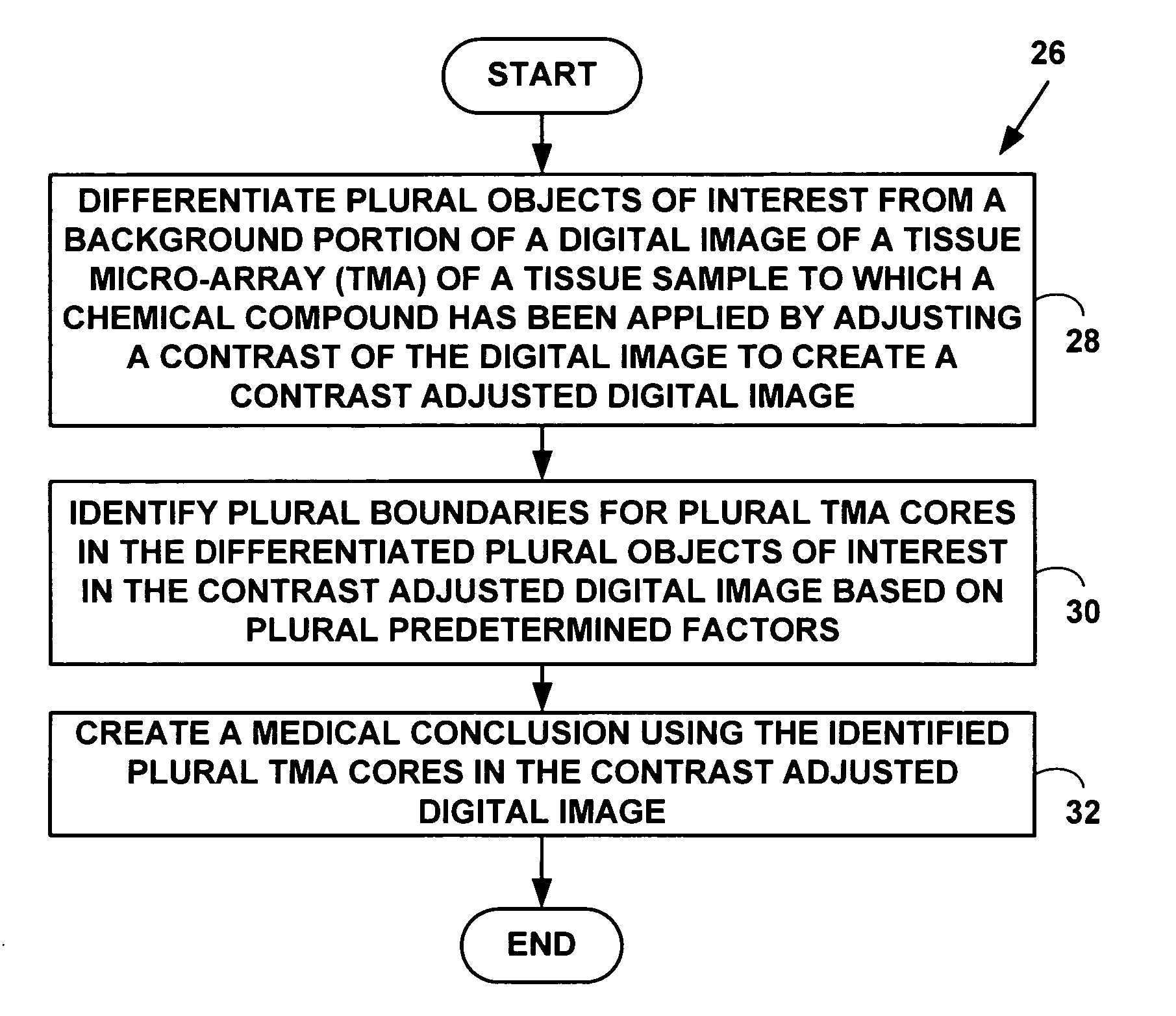
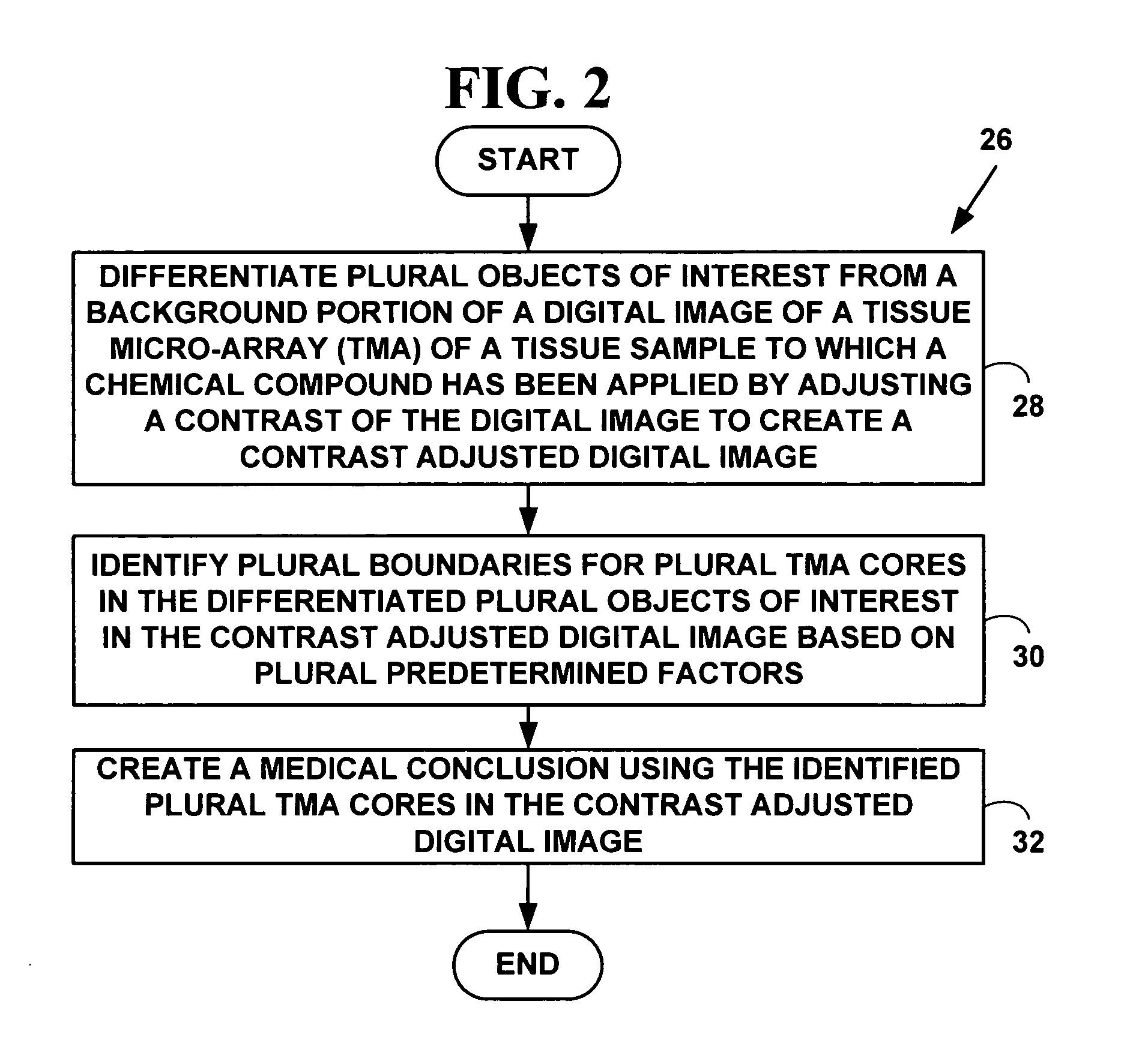
Embodiment Construction

Exemplary Automated Biological Sample Analysis System

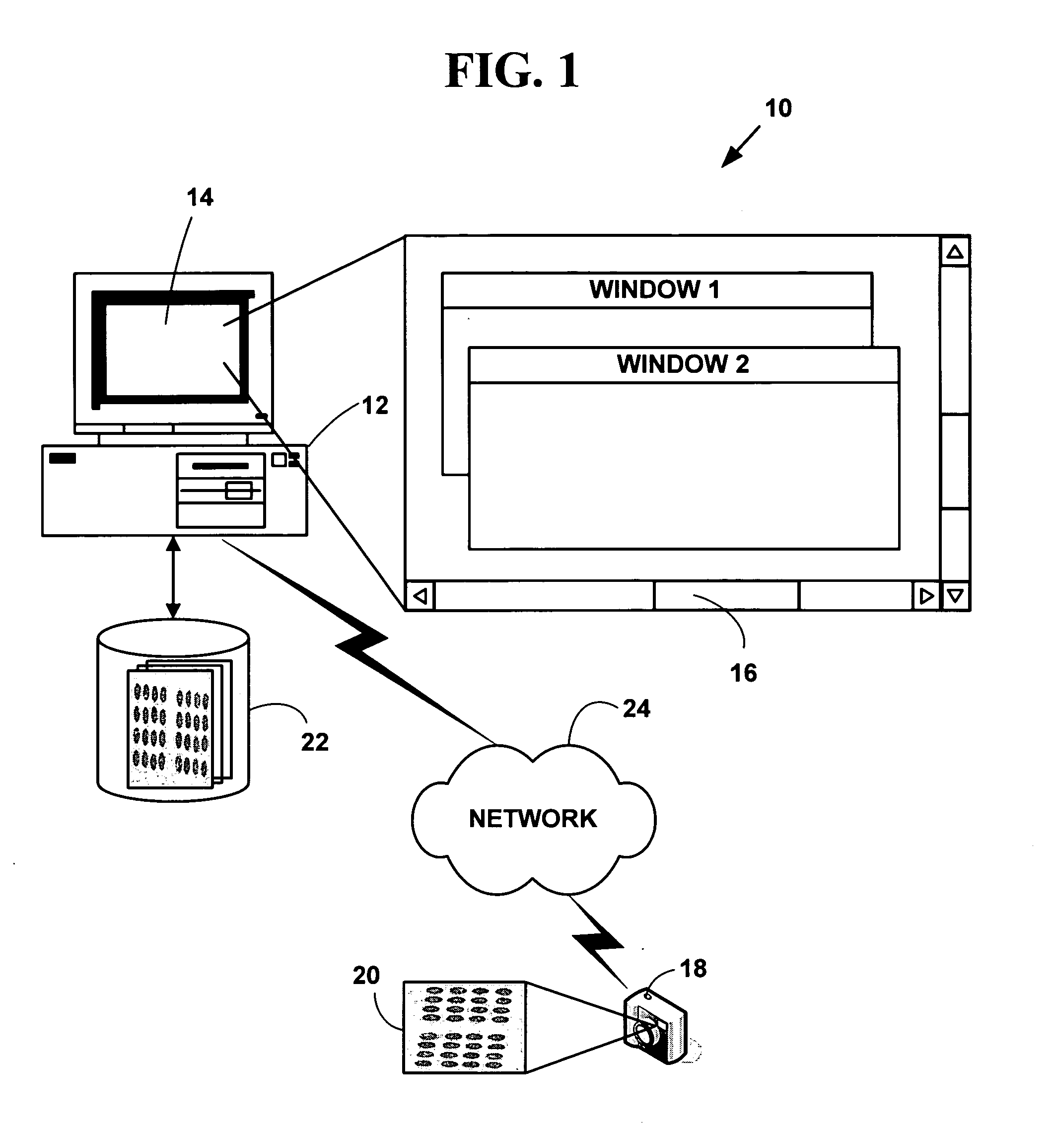
[0043]FIG. 1 is a block diagram illustrating an exemplary automated biological sample processing system 10. The exemplary system 10 includes one or more computers 12 with a computer display 14 (one of which is illustrated). The computer display 14 presents a windowed graphical user interface (“GUI”) 16 with multiple windows to a user. The system 10 may optionally include a microscope or other magnifying device (not illustrated in FIG. 1). The system 10 further includes a digital camera 18 (or analog camera) used to provide plural digital images 20 in various digital images or digital data formats. One or more databases 22 (one or which is illustrated) include biological sample information in various digital images or digital data formats. The databases 22 may be integral to a memory system on the computer 12 or in secondary storage such as a hard disk, floppy disk, optical disk, or other non-volatile mass storage devices. The co...


PUM

PropertyMeasurementUnit
Sizeaaaaaaaaaa
Sizeaaaaaaaaaa
Lengthaaaaaaaaaa

Abstract

A method and system for automated quantitation of tissue micro-array image (TMA) digital analysis. The method and system automatically analyze a digital image of a TMA with plural TMA cores created using a needle to biopsy or other techniques to create standard histologic sections and placing the resulting needle cores into TMA. The automated analysis allows a medical conclusion such as a medical diagnosis or medical prognosis (e.g., for a human cancer) to be automatically determined. The method and system provides reliable automatic TMA core gridding and automated TMA core boundary detection including detection of overlapping or touching TMA cores on a grid.

Description

CROSS REFERENCES TO RELATED APPLICATIONS [0001] This application claims priority to U.S. Provisional Patent Application No. 60 / 573,262 filed May 21, 2004, the contents of which are incorporated by reference. This application claims also priority to U.S. patent application Ser. No. 10 / 938,314, filed Sep. 10, 2004, which claims priority U.S. Provisional Patent Application No. 60 / 501,142, filed Sep. 10, 2003, and U.S. Provisional Patent Application No. 60 / 515,582 filed Oct. 30, 2003, and this application claims also priority to U.S. patent application Ser. No. 10 / 966,071, filed Oct. 15, 2004 which claims priority to U.S. Provisional Patent Application No. 60 / 530,714, filed Dec. 18, 2003, the contents of all of which are incorporated by reference.COPYRIGHT NOTICE [0002] Pursuant to 37 C.F.R. 1.71(e), applicants note that a portion of this disclosure contains material that is subject to and for which is claimed copyright protection, such as, but not limited to, digital photographs, scree...

Claims


Application Information

Patent Timeline
IPC IPC(8): G01N33/48G06K9/00G06F19/00
CPCG06K9/00127G06V20/69
Inventor GHOLAP, ABHIJEET S.GHOLAP, GAURI A.JADHAV, PRITHVIRAJBARSKY, SANFORD H.RAO, C. V. K.VIPRA, MADHURA
Owner VENTANA MEDICAL SYST INC
Why Eureka
Try Eureka