Intubation and imaging device and system

a technology of imaging device and intubation tube, which is applied in the field of endotracheal intubation tool, can solve the problems of difficult visualization of posterior pharyngeal area, difficulty in intubation, and difficulty in performing intubation

Inactive Publication Date: 2006-01-26
GIVEN IMAGING LTD
View PDF20 Cites 85 Cited by
  • Summary
  • Abstract
  • Description
  • Claims
  • Application Information

AI Technical Summary

Benefits of technology

[0006] Some embodiments of the present invention include endotracheal intubation tools such as laryngoscopes, and may include for example an imaging unit, which allows typically continuous in vivo visualization during insertion, and possibly during use, of the laryngoscope. Endotracheal intubation may be a life saving procedure performed in emergency situations, such as in the case of an obstructed airway. Thus, the p

Problems solved by technology

In many patients, intubation may be particularly difficult to perform due to morphological anomalies, such as a large tongue, excessive soft tissue, or tracheal displacement.
These morphological anomalies may make it difficult to visualize the posterior pharyngeal area, larynx and cords, and may cause difficulties in intubation.
In medical emergency situations, an attempt to i

Method used


Image

  • Intubation and imaging device and system
  • Intubation and imaging device and system
  • Intubation and imaging device and system

Examples


Embodiment Construction

[0016] In the following description, various aspects of the present invention will be described. For purposes of explanation, specific configurations and details are set forth in order to provide a thorough understanding of the present invention. However, it will also be apparent to one skilled in the art that the present invention may be practiced without the specific details presented herein. Furthermore, well known features may be omitted or simplified in order not to obscure the present invention.

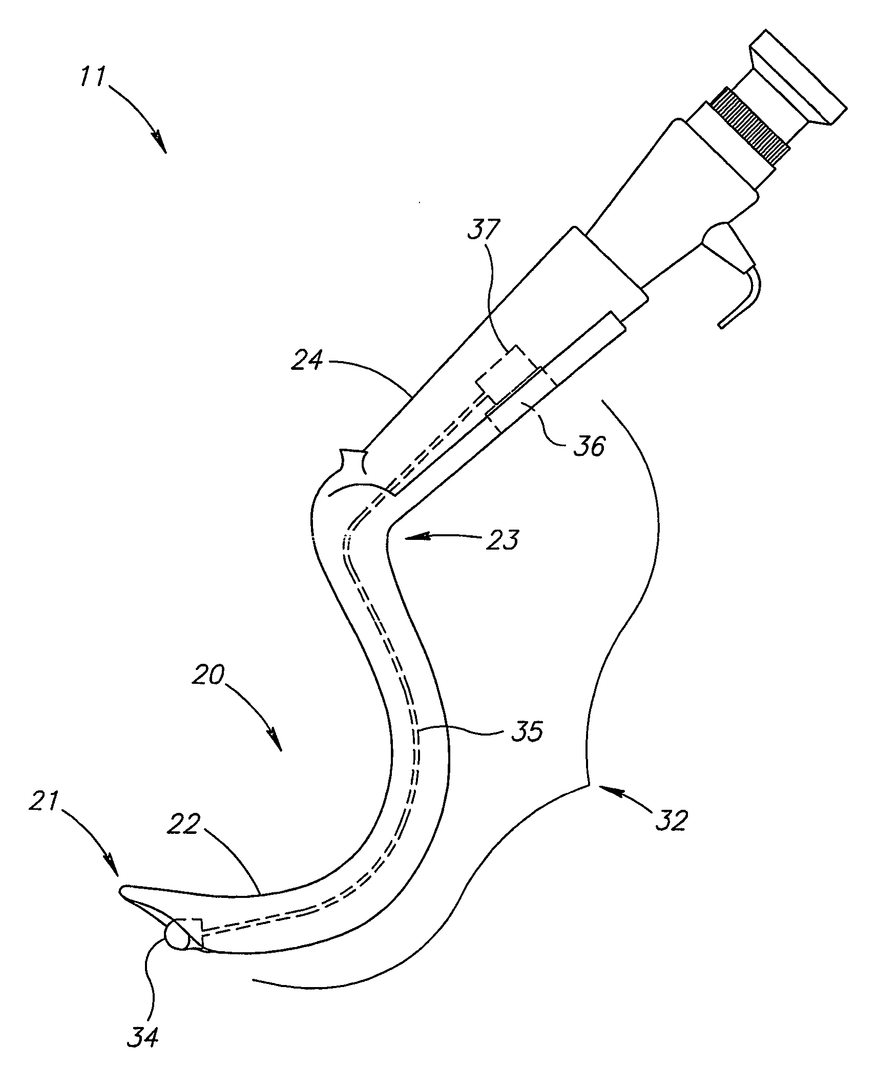
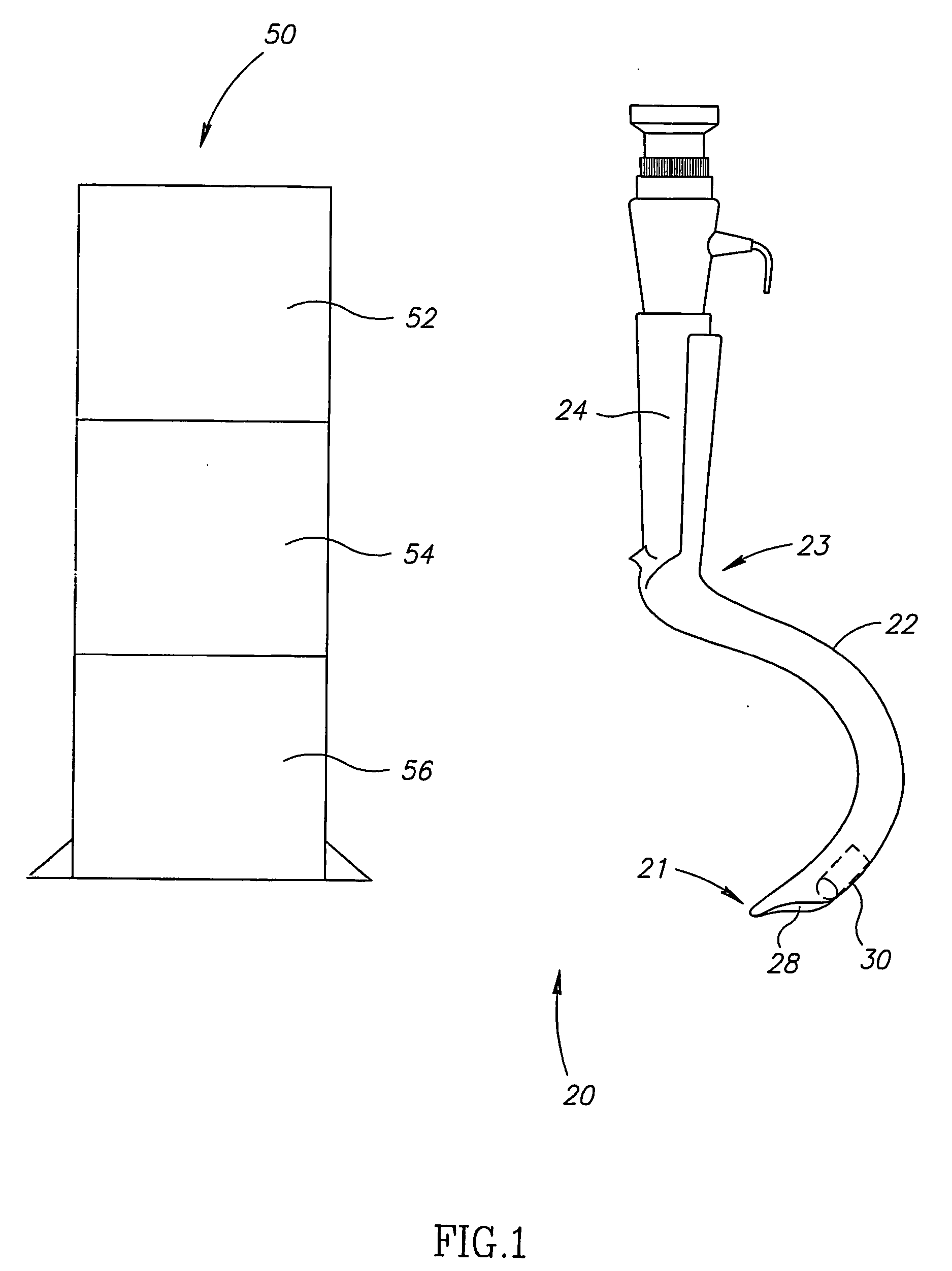
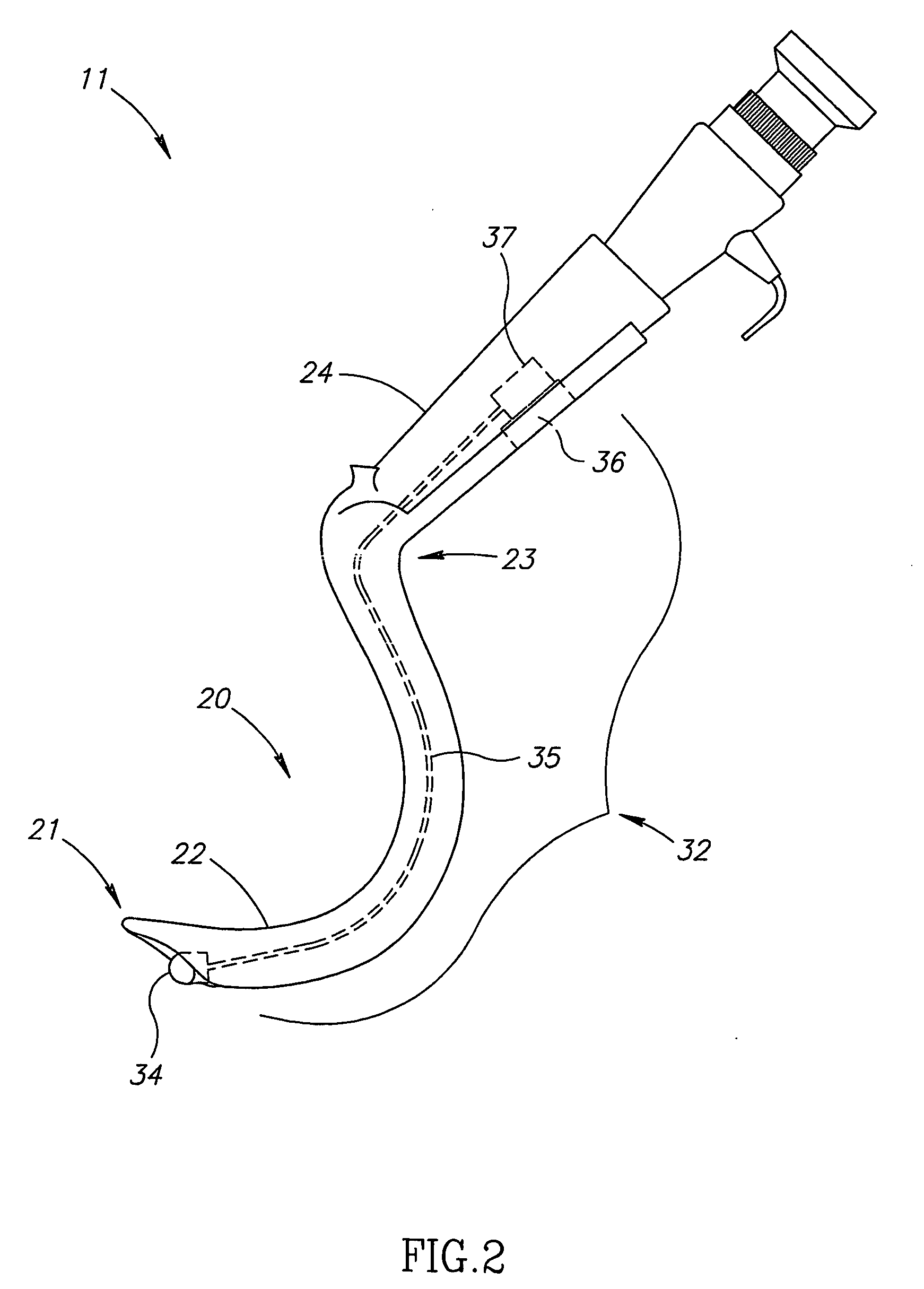
[0017] Reference is now made to FIG. 1, which is a schematic illustration of an intubation system 10 in accordance with an embodiment of the invention. The intubation system 10 typically includes a laryngoscope 20, an imaging unit 30 attached to or included as part of laryngoscope 20 and a receiving unit 50. receiving unit 50, which, according to one embodiment includes a receiver 52, a processor 54 and a screen or display 56, receives signals, for example image data, from imaging unit...


PUM


Abstract

An intubation tool may include an imaging unit. The imaging unit may be, for example, self contained, autonomous and/or single use. Images may be transmitted to, for example, an external receiving system.

Description

FIELD OF THE INVENTION [0001] The present invention relates to the field of medical devices, more specifically to an endotracheal intubation tool having integrated visualization and to a method for its use. BACKGROUND OF THE INVENTION [0002] Endotracheal intubation is a common medical procedure often directed at opening a closed larynx by inserting a laryngoscope through the larynx followed by the insertion of an endotracheal tube, which enables air supply to the patient. Endotracheal intubation is a typically life saving procedure performed in emergency cases. Thus, the ability to intubate a patient rapidly is highly important. [0003] In many patients, intubation may be particularly difficult to perform due to morphological anomalies, such as a large tongue, excessive soft tissue, or tracheal displacement. These morphological anomalies may make it difficult to visualize the posterior pharyngeal area, larynx and cords, and may cause difficulties in intubation. In medical emergency s...

Claims


Application Information

Patent Timeline
IPC IPC(8): A61B1/267A61B1/06A61B1/05A61B5/00A61B19/00A61M
CPCA61B1/00016A61B1/00105A61B1/041A61B5/0013A61B1/267A61B1/0676A61B1/05
Inventor GILREATH, MARK G.
Owner GIVEN IMAGING LTD
Why Eureka
Try Eureka