Optimizing image signal interpretation for analog image signals from medical image recording devices

an image signal and image signal technology, applied in image data processing, medical science, instruments, etc., can solve problems affecting the correlation of image data and position data

Inactive Publication Date: 2006-02-02
BRAINLAB
View PDF6 Cites 2 Cited by
  • Summary
  • Abstract
  • Description
  • Claims
  • Application Information

AI Technical Summary

Benefits of technology

[0005] The present invention provides a method for optimizing the interpretation of analog image signals or sequences of image signals output by medical image recording devices, such as x-ray apparatus, that overcomes one or more of the disadvantages discussed above. In particular, the invention enables one to ascertain the moment when a new image signal is detected without additional or dedicated hardware. Further, the invention enables the type and properties of new images to be interpreted.
[0007] In other words, the present invention enables one to move away from using hardware to detect the moment a new image is produced. More specifically, the present invention can interpret the image signal and deduce from the contents of the image signal or sequence of image signals whether a new image has been produced. This is advantageous in that less hardware is required to operate the system. Further, in interpreting the detected image signals, additional information can be obtained that can be used during surgery.
[0008] As will be discussed below, the threshold value preferably is dynamically adjusted. If the threshold value were not adjusted, instances could arise where a new recorded image that does not change over a certain period of time, constantly lies below the threshold value due to an increased proportion of noise, thus affecting the correlation of the image data and the position data. A system that does not adapt the threshold value would constantly output a signal indicating new images are being detected. By adapting the threshold value, this source of error can be eliminated.
[0010] As already mentioned above, constantly analyzing the image contents allows more than just establishing whether a new image is pending at the image receiver of an analog x-ray apparatus. It also enables the identification of whether or not only particular recording parameters have changed or whether the end of a long-term x-ray recording has been reached.

Problems solved by technology

If the threshold value were not adjusted, instances could arise where a new recorded image that does not change over a certain period of time, constantly lies below the threshold value due to an increased proportion of noise, thus affecting the correlation of the image data and the position data.

Method used


Image

  • Optimizing image signal interpretation for analog image signals from medical image recording devices
  • Optimizing image signal interpretation for analog image signals from medical image recording devices
  • Optimizing image signal interpretation for analog image signals from medical image recording devices

Examples


Embodiment Construction

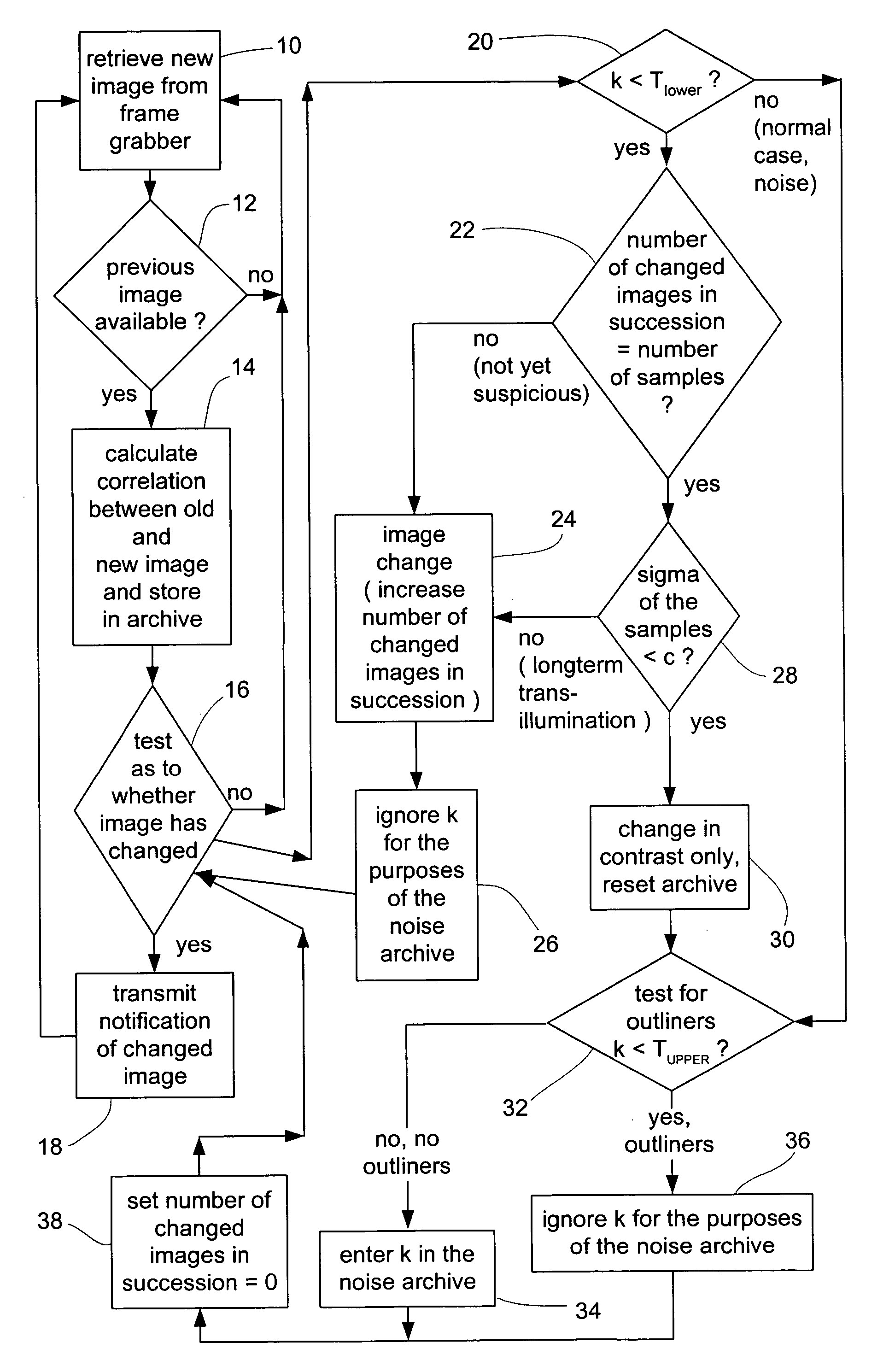
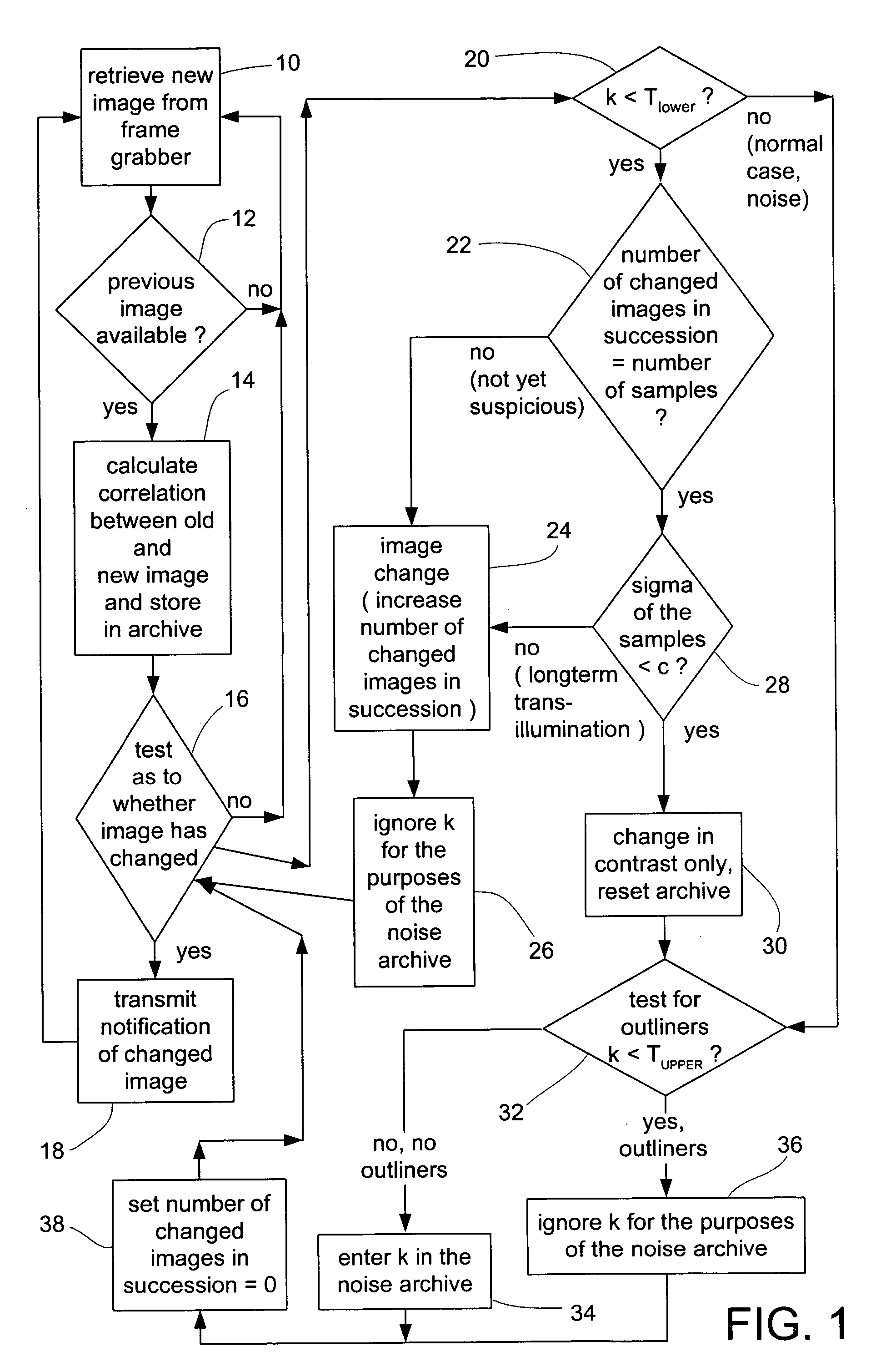
[0023] The invention will now be described with reference to the drawings. It is noted that the features of the invention can be implemented individually or in any combination. Further, the invention can be used with various medical image recording devices, including, for example, computer or nuclear spin tomographs or the like. As used herein, the term “adjust(ing) the threshold value” refers to setting, resetting, adapting, changing, etc., the threshold value based on certain criteria.

[0024] In general terms, the invention can be classified as an evaluation of an auto-correlation value for a medical imaging system (e.g., a C-arc x-ray apparatus) video output signal. More specifically, the auto-correlation value or correlation is calculated by comparing sequential frames of the video signal, wherein the correlation range is less than or equal to 1.0 (perfect correlation, no changes between sequential frames) and greater than or equal to −1.0 (no correlation between sequential fram...


PUM


Abstract

A method for optimizing the interpretation of analog image signals or sequences of image signals output by medical image recording devices. The correlation of consecutively recorded image signals is tested and, based on the test, the image signals are identified as depicting the same or different images. More specifically, if the correlation is not less than a particular threshold value, it is established that the image signals depict the same image. If the correlation is less than a particular threshold value, it is established that the image signals possibly depict different images. Further, the threshold value is dynamically adjusted if the correlation has changed.

Description

RELATED APPLICATION DATA [0001] This application claims priority of U.S. Provisional Application No. 60 / 606,008 filed on Aug. 31, 2004, which is incorporated herein by reference in its entirety.FIELD OF THE INVENTION [0002] The invention relates to a method for optimizing the interpretation of analog image signals or sequences of image signals output by medical image recording devices. BACKGROUND OF THE INVENTION [0003] In the field of x-ray image-assisted, image-guided surgery a physician works with the aid of a navigation system that provides information on the inner body structure of a patient with the aid of intraoperatively produced x-ray images. In such image guided surgery, it is important to precisely identify the moment at which a new image is recorded by the x-ray apparatus. The navigation system has to “know” the moment when the x-ray apparatus recorded a particular x-ray image in order to correctly assign said x-ray image information to corresponding patient position / loc...

Claims


Application Information

Patent Timeline
Patent Type & Authority Applications(United States)
IPC IPC(8): G06K9/00
CPCG06T7/0002G06T2207/30004G06T2207/10116Y10S128/922
Inventor KRAUS, FLORIANMAIER, CHRISTIANWEISER, MANFRED
Owner BRAINLAB
Why Eureka
Try Eureka