
Minimally invasive medical implant and insertion device and method for using the same

a medical implant and minimally invasive technology, applied in the field of minimally invasive medical implants, can solve the problems of increased post-operative pain and/or infection risk to at least a small degree, the use of needles to pass the tape through the body poses a risk of vessel, bladder and bowel perforation,

Active Publication Date: 2006-02-02
ETHICON INC
View PDF85 Cites 107 Cited by
  • Summary
  • Abstract
  • Description
  • Claims
  • Application Information

AI Technical Summary

Benefits of technology

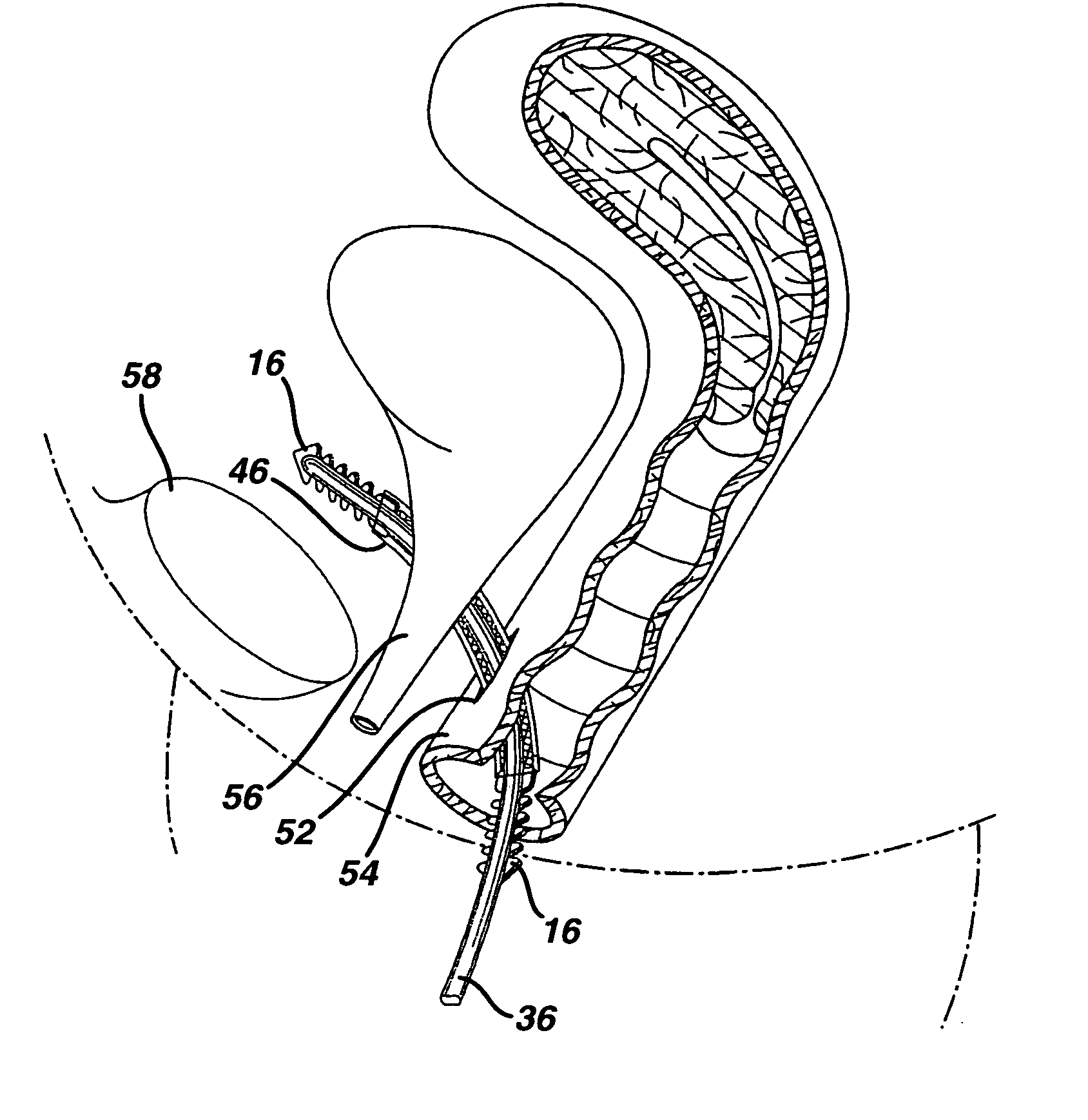
[0010] The present invention provides a medical implant inserter including an inserter device having a distal end region including a tissue penetrating distal tip, a proximal end region, a capture element extending outwardly therefrom, and a first pass through element extending outwardly therefrom and having at least one opening therethrough, with the pass through element being positioned at a location proximal of the capture element. The inserter device further includes an implant holding element having a proximal end and a distal end. The implant holding element is removably coupled at its distal end to the inserter device by being removably received within the capture element, and is movably coupled to the inserter device at a location proximal of the capture element. A portion of the implant holding element lies substantially adjacent to the inserter device, and a distal end portion extends to a position wherein it is spaced apart from the inserter device before being removably received within the capture element. The implant holding element passes through the at least one opening in the first pass through element to thereby at least partially maintain the position of the implant holding element relative to the inserter device.
[0011] A medical device is also provided including an implant for implantation within a patient, and a first inserter for inserting the implant. The first inserter has a distal end region including a tissue penetrating distal tip, a proximal end region, a capture element located at the distal end region, and an implant holding element having a proximal end and a distal end, the distal end of which is removably received within the capture element. The implant holding element further is movably coupled to the first inserter at a first location proximal of the capture element. The implant holding element extends from the first location at which it is movably coupled to the first inserter, and subsequently through the implant before being removably received within the capture element to thereby removably secure the implant to the insertion device.
[0013] Finally, a method for implanting a suburethral implant is provided including the steps of providing an implant including an implantable, elongated tape having a multiplicity of openings formed therethrough, the tape having a first end region and a second end region longitudinally opposite the first end region, and first and second bio-compatible fixation elements attached to the first and second end regions respectively, and providing first and second insertion devices each including a first inserter having a distal end region including a tissue penetrating distal tip, a proximal end region, a capture element located at the distal end region, and an implant holding element having a proximal end and a distal end. The method further includes removably coupling the first and second fixation elements of the implant to the first and second inserters respectively by extending the respective implant holding element from a first location wherein it is movably coupled to the insertion device, through the fixation element and to a second location wherein its distal end is removably received within the capture element, inserting the first inserter and attached first fixation element through a vaginal incision and into a patient's tissue on a first side of the urethra, inserting the second inserter and attached second fixation element through the vaginal incision and into the patient's tissue on a second side of the urethra, adjusting the first inserter and attached fixation element and the second inserter and attached fixation element to thereby properly position the implant to provide support to the patient's urthra, uncoupling the first and second inserters from the first and second fixation elements substantially without further manipulation of the implant, and leaving the implant implanted within the body without further adjustment thereof.

Problems solved by technology

As a result of this condition, involuntary urine leakage occurs from simple physical activity, such as running or jumping, and even coughing or sneezing, as the urethra is not properly supported and does not remain fully closed during such activity.
One of the few disadvantages of known procedures for implanting sub-urethral tapes is that the use of needles to pass the tape through the body poses a risk for vessel, bladder and bowel perforation.
This, of course, increases the risk of post-operative pain and / or infection to at least a small degree.

Method used


Image

  • Minimally invasive medical implant and insertion device and method for using the same
  • Minimally invasive medical implant and insertion device and method for using the same
  • Minimally invasive medical implant and insertion device and method for using the same

Examples


Embodiment Construction

[0036] Although the present invention is described in detail in relation to its use as a sub-urethral tape for treating stress urinary incontinence, it is to be understood that the invention is not so limited, as there are numerous other applications suitable for such an implant. For example, implants incorporating novel features described herein could be used for repairing pelvic floor defects such as, but not limited to, cystoceles and rectoceles, and for hernia repair or other prolapse conditions, or for supporting or otherwise restoring other types of tissue.

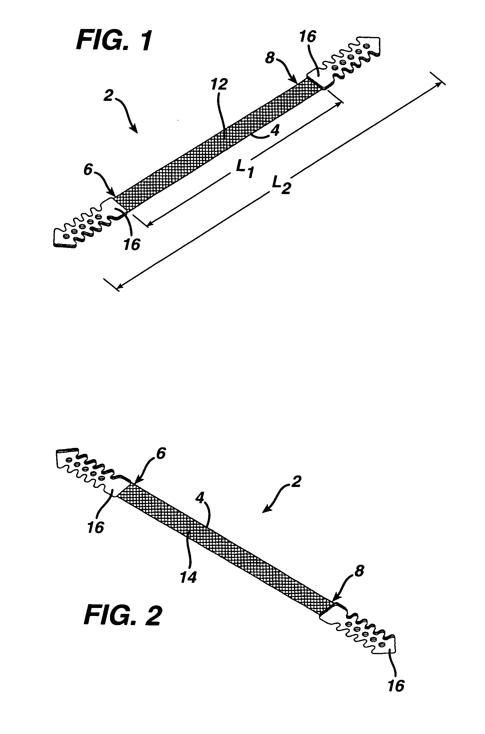
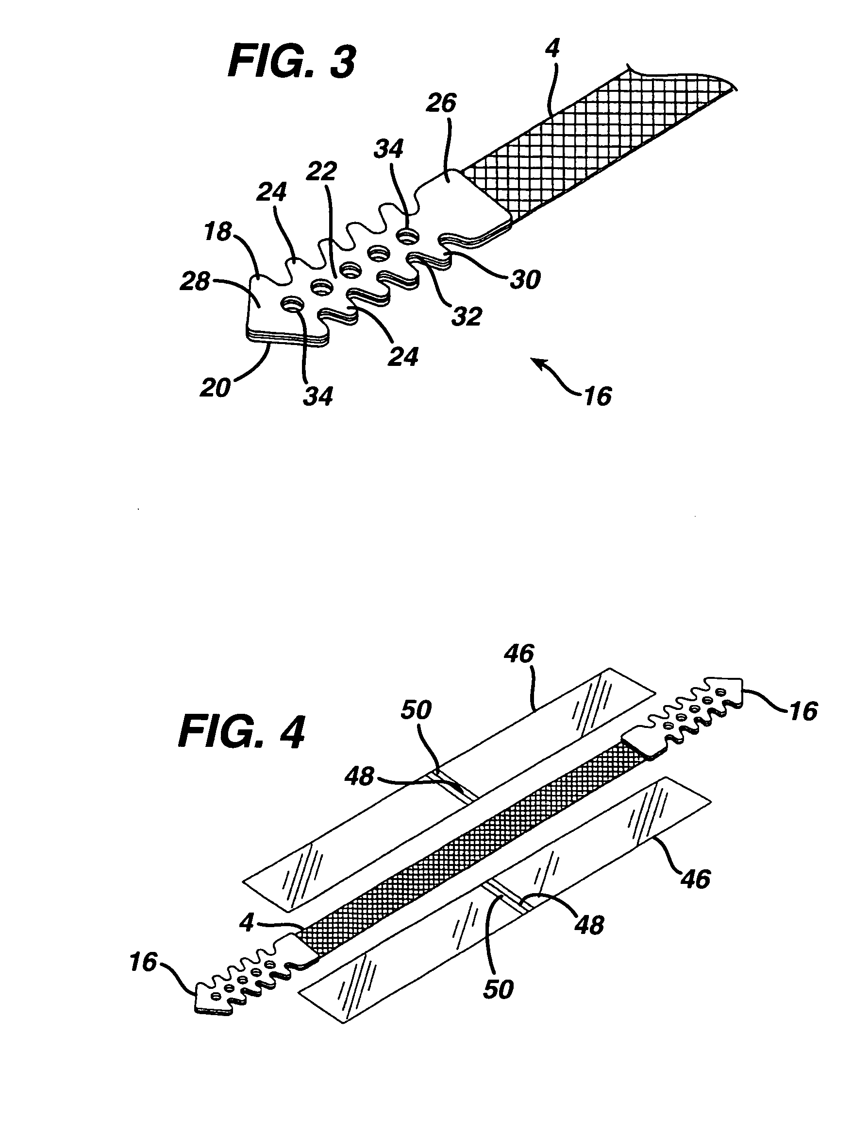
[0037] Turning initially to FIGS. 1-3 of the drawings, one embodiment of an implant 2 in the form of a sub-urethral tape particularly suited for the treatment of stress urinary incontinence (SUI) includes an implantable, elongated tape 4. The main tape portion 4 has a multiplicity of openings formed through the thickness thereof, and includes a first end 6 and a second end 8 longitudinally opposite the first end 6.

[0038] P...


PUM


Abstract

A medical device including an implant and inserter and a method for using the same. One embodiment includes an implant for implantation within a patient, and a first inserter for inserting the implant. The first inserter has a distal end region including a tissue penetrating distal tip, a proximal end region, a capture element located at the distal end region, and an implant holding element having a proximal end and a distal end. The distal end is removably received within the capture element, and the implant holding element further is movably coupled to the first inserter at a first location proximal of the capture element. The implant holding element extends from the first location at which it is movably coupled to the first inserter, and subsequently through the implant before being removably received within the capture element to thereby removably secure the implant to the insertion device.

Description

CROSS REFERENCE TO RELATED APPLICATIONS [0001] The present application claims priority to U.S. Provisional Patent Application Ser. Nos. 60 / 591,648, filed on Jul. 28, 2004, and ______, filed on Jul. 20, 2005.BACKGROUND OF THE INVENTION [0002] 1. Field of the Invention [0003] This invention relates generally to minimally invasive medical implants and procedures for their use and insertion device for implanting the same, having particular application for treating urinary incontinence. [0004] 2. Description of the Prior Art [0005] Stress urinary incontinence (SUI) is a female medical condition commonly associated with weakening of the pelvic muscles and / or connective tissues that support the urethra in its proper position. As a result of this condition, involuntary urine leakage occurs from simple physical activity, such as running or jumping, and even coughing or sneezing, as the urethra is not properly supported and does not remain fully closed during such activity. [0006] A widely ac...

Claims


Application Information

Patent Timeline
IPC IPC(8): A61B17/10
CPCA61B17/06066A61B17/06109A61F2/0045A61B2017/00805A61B2017/06042A61B17/062A61F2250/0018
Inventor SMITH, DANIEL J.NORDMEYER, MICHAELSUMP, RAIMOLANDGREBE, SUSANNEPETERS, BURKHARD
Owner ETHICON INC
Learn More