Ultra-fine micropsy needle

Inactive Publication Date: 2006-03-09
BRAUTIGAM ROBERT T +2
View PDF6 Cites 12 Cited by
  • Summary
  • Abstract
  • Description
  • Claims
  • Application Information

AI Technical Summary

Benefits of technology

[0005] A further object of this invention is to provide such an ultra-fine needle which can be easily manipulated for retrieving a specimen.
[0006] A goal of this invention is the development of an ultra-fine biopsy needle that: 1) may penetrate the skin between nerve endings and therefore be painless, and 2) may inflict minimal tissue trauma because of the small diameter and small size of the sample removed, thus dramatically reducing the risk of hemo

Problems solved by technology

This technique employs 18-2 OGa needles, but samples are inadequate for conventional histologic analysis and provide only cytological information.
Currently the major limitations to these biopsy techniques are the associated tissue injury and associated complications.
Complications include pain during the procedure, bleeding, organ damage and subseque

Method used


Image

  • Ultra-fine micropsy needle
  • Ultra-fine micropsy needle
  • Ultra-fine micropsy needle

Examples


Embodiment Construction

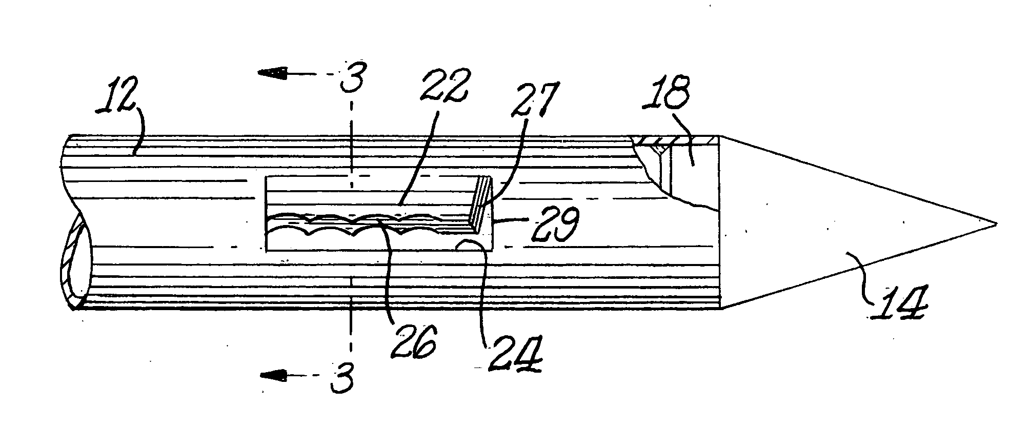
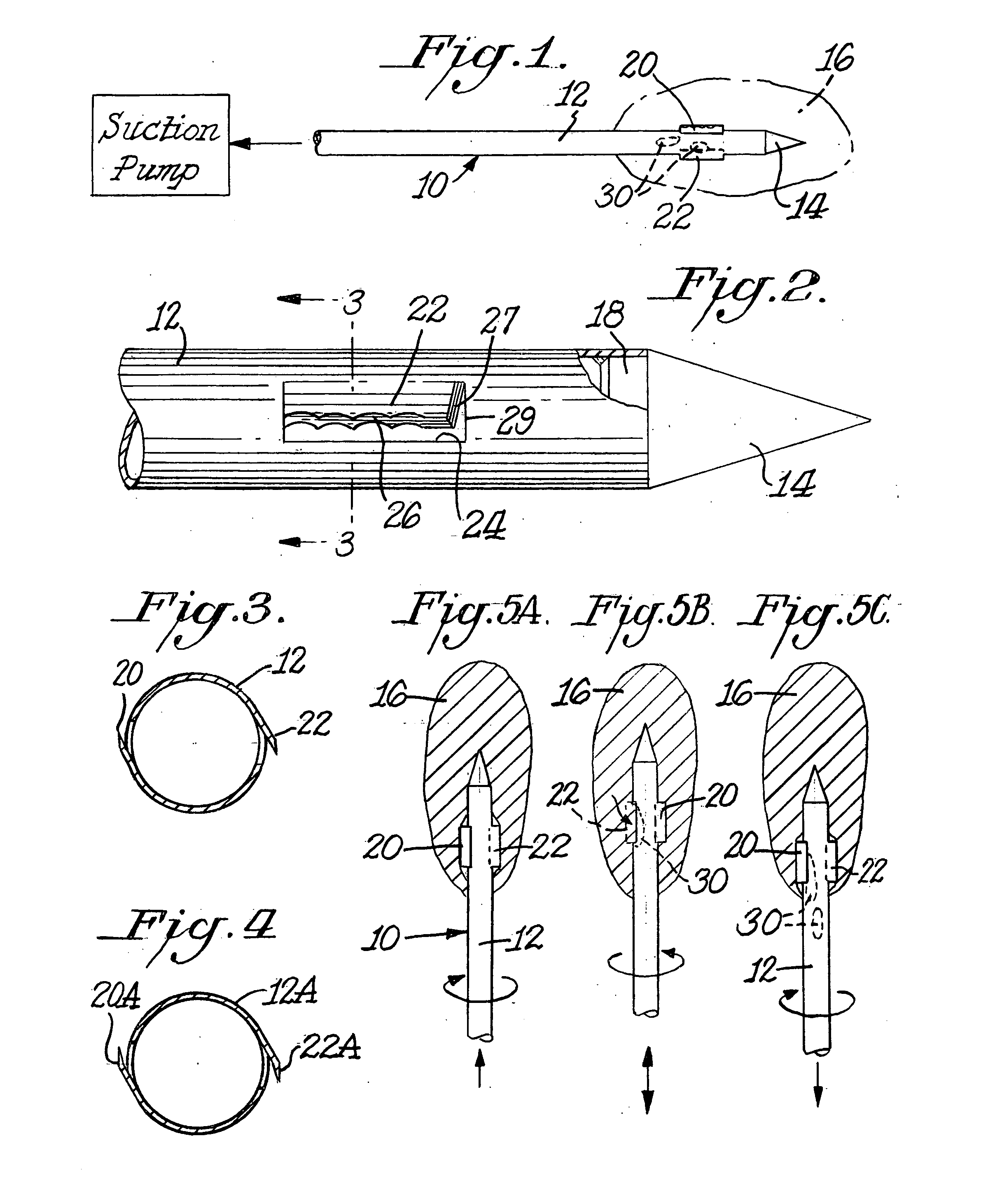
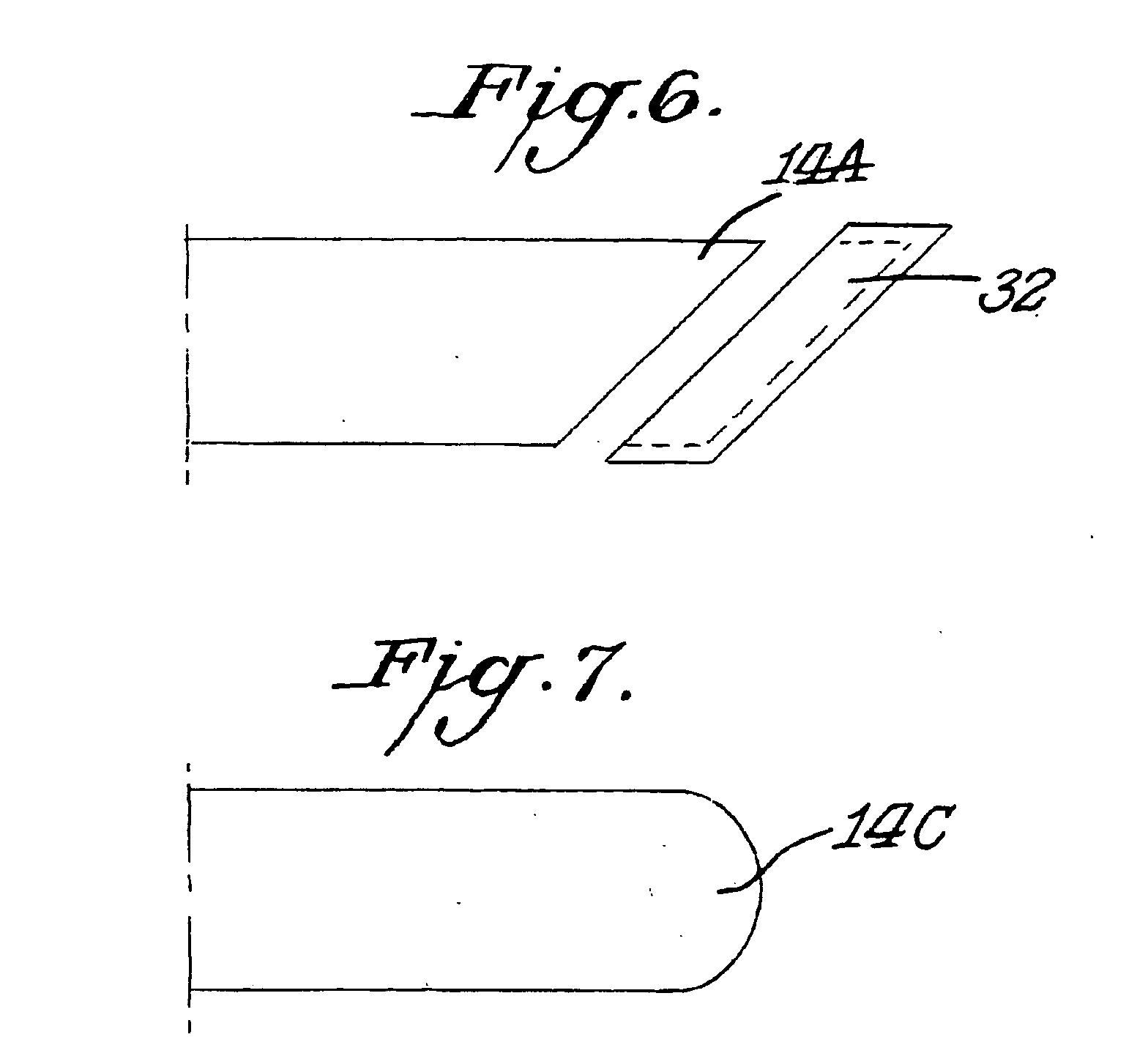
[0017] The present invention is directed to providing an ultrafine micropsy needle which would dramatically alter the risk / benefit ratio when it comes to the percutaneous sampling of human tissues for diagnostic purposes. In accordance with this invention the ultra-fine micropsy needle has an outside diameter of less than 1.0 mm and preferably in the range of 0.01 mm to less than 1.0 mm. The maximum outside diameter could be no greater than 0.8 mm or no greater than 0.6 mm or no greater than 0.4 mm or no greater than 0.2 mm or no greater than 0.1 mm.

[0018] In order to provide such a needle proper manufacturing techniques and materials would have to be used to attain the functional designs of the desired dimensions.

[0019] The invention could be practiced where the needle is a specimen retrieving needle having structure for cutting and then retrieving a specimen.

[0020] In accordance with this invention a specimen retrieving needle is provided which is may be considered as an ultra-...


PUM


Abstract

A specimen retrieving needle is in the form of an elongated body having a closed lead end. At least one cutting blade may be formed on the body. The blade may have a sharpened edge so as to remove a specimen from an internal organ. The needle is ultrathin with an outside diameter of less than 1.0 mm to minimize tissue trauma and to reduce the risk of pain, hemorrhage and organ damage.

Description

BACKGROUND OF THE INVENTION [0001] Conventional biopsy needles and devices are designed to retrieve a sample or specimen that has an adequate size required for conventional histologic, cytologic or protein analyses. The actual size of current devices is ultimately dictated by the tissue and disease process that is the center of investigation. Generally, the size is not less than 14-16 Ga (0.065-0.083 inches OD or 0.047-0.063 inches ID). Smaller tissue samples can be obtained through a process of fine-needle aspiration. This technique employs 18-2 OGa needles, but samples are inadequate for conventional histologic analysis and provide only cytological information. Currently the major limitations to these biopsy techniques are the associated tissue injury and associated complications. Complications include pain during the procedure, bleeding, organ damage and subsequent infection. Clinicians constantly must weigh the risk of the biopsy procedure with potential benefits to the patient....

Claims


Application Information

Patent Timeline
IPC IPC(8): A61B10/00A61B17/32
CPCA61B10/0266A61B2017/00345A61B10/0283
Inventor BRAUTIGAM, ROBERT T.PERDRIZET, GEORGE A.BRAUTIGAM, RICHARD H.
Owner BRAUTIGAM ROBERT T
Why Eureka
Try Eureka