Enhancement magnetizer for magnetic resonance imaging screening

a magnetic resonance imaging and enhancement magnet technology, applied in the field of enhancement magnetizers for magnetic resonance imaging screening, can solve the problems of increasing the risk of magnetic resonance imaging, increasing the risk of damage to the mri magnet itself, and achieving the effect of convenient and convenient application

Inactive Publication Date: 2006-04-20
MEDNOVUS
View PDF39 Cites 18 Cited by
  • Summary
  • Abstract
  • Description
  • Claims
  • Application Information

AI Technical Summary

Benefits of technology

[0023] The second fundamental advantage of the present invention is that magnetization can occur in all three (x, y, and z) axes. This is accomplished according to the present invention by positioning the magnetizer appropriately on all three axes around a ferromagnetic threat object. This can be done with a hand held magnet. Where scanning of a person's head is desired, to detect a threat object such as a bobby pin, for instance, magnetization can be achieved according to the present invention by placing the subject's head into a helmet-like structure. In this embodiment, the helmet can incorporate magnetic means, and the method of applying the helmet can be designed to magnetize any threat object in three axes. In either embodiment, the method and apparatus of the present invention solve the problem of magnetizing in only one axis, as a magnetic field can be easily and readily applied in all three axes.
[0024] The novel features of this invention, as well as the invention itself, will be best understood from the attached drawings, taken along with the following description, in which similar reference characters refer to similar parts, and in which:
[0025]FIG. 1 is a graph of the initial magnetization curve of a bobby pin;
[0026]FIG. 2 is a schematic view of a magnetic field generated by a permanent magnet;
[0027]FIGS. 3A and 3B are elevation views of a first embodiment of hand held magnetizers according to the present invention;
[0028]FIGS. 4A and 4B are elevation views of a second embodiment of hand held magnetizers according to the present invention;

Problems solved by technology

Magnetic resonance imaging has significant risks, however, and these are becoming more apparent as the number of MRI procedures increases dramatically.
In particular, ferromagnetic objects are drawn toward the magnetic resonance imaging magnet by the strong magnetic field of the magnet, sometimes with catastrophic results.
This attraction of ferromagnetic objects to the MRI magnet is termed the “missile threat.” Not only have there been numerous injuries to patients, including one tragic death, but damage to the MRI magnet itself is also a significant problem.
The huge number of false positive alarms generated by conventional metal detectors has caused such consternation for MRI staff technicians that conventional metal detectors have been abandoned for this application.
Indeed, conventional metal detectors may have no current usefulness as a practical solution for MRI safety.
A tragic death has occurred from the missile threat, and many “near misses” have occurred.
It is well known that very serious accidents can occur if ferromagnetic objects are carried into the vicinity of an MRI scanner.
So, tempered steel or other magnetically hard objects like bobby pins are just as dangerous as ordinary soft steel items, from the missile threat point of view, and it is important that these be detected.
Some types of magnetic fields are insufficient to magnetize certain ferromagnetic threat objects, including bobby pins and the like, for detection by a ferromagnetic-detecting portal or a ferromagnetic-detecting wand.
In addition, the earth's magnetic field for a particular location on the planet is in only one axis, and detection can be missed when the major axis of a ferromagnetic threat is perpendicular to the earth's magnetic field.
This means that a bobby pin would be very poorly detected, if at all, by a passive portal that depended on the earth's field to magnetize the objects being detected.
In addition, the MRI fringing field does not apply magnetization in all three axes, and, if the threat object is perpendicular to the fringing magnetic field, detection is often compromised because of insufficient magnetization.
As a final example, the permanent magnets or coil arrangements of a strength typically found on an “active” portal or wand may be insufficient to magnetize certain difficult-to-magnetize ferromagnetic objects, such as bobby pins.
This can result in insufficient magnetization, especially of difficult-to-magnetize ferromagnetic threat objects.
For instance, if an applied field is only applied to provide detection of a threat for the x axis, it is possible that a threat may escape detection because of insufficient magnetization for the y axis and the z axis.
Further, the chance of detecting a threat object in such a portal is increased if the subject is asked to rotate or tilt within the portal pass-through aperture.
There may be additional reasons for insufficient magnetization to support detection of a threat object.
The object may be tiny, or the configuration of the object may make magnetization, and hence, detection, difficult.
For instance, a round object is more difficult to magnetize and detect than an elongated one.
Or, the threat object may be located at too great a distance from a ferromagnetic-detecting wand, such as when the wand is waved at too great a distance from the threat object to achieve the required magnetization.
In any of the above instances, as well as others, the result is insufficient magnetization to allow the sensing system of the portal or wand apparatus to detect the threat, resulting in a false negative (missed) response.

Method used


Image

  • Enhancement magnetizer for magnetic resonance imaging screening
  • Enhancement magnetizer for magnetic resonance imaging screening
  • Enhancement magnetizer for magnetic resonance imaging screening

Examples


first embodiment

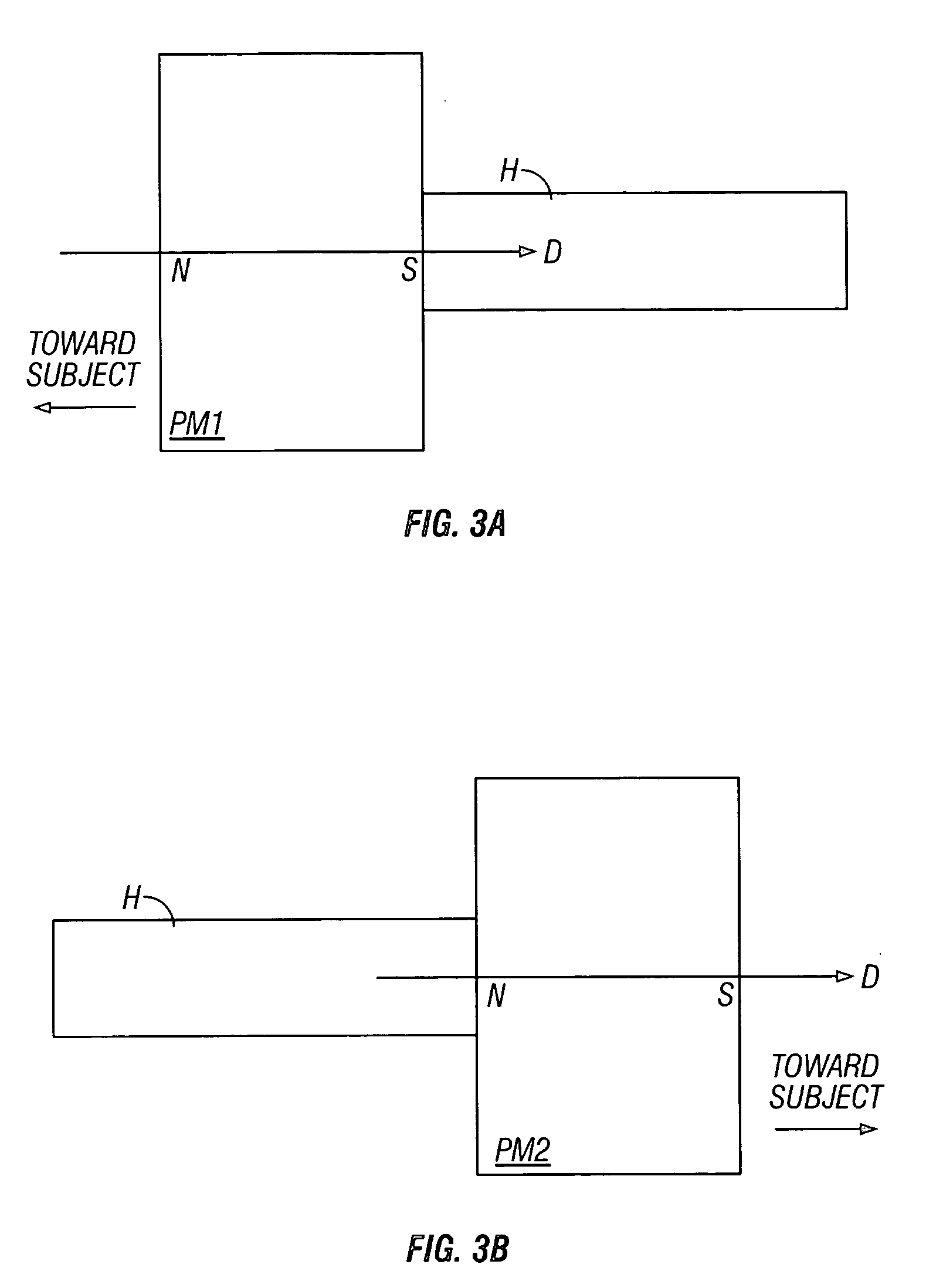
[0033] Preferably, for ease of use, the hand-held permanent magnet is attached to a non-ferromagnetic handle or, alternatively, to a non-ferromagnetic strap. FIGS. 3A and 3B show the apparatus of the present invention, namely a pair of hand-held permanent magnets, each with a non-ferromagnetic handle H, made of a material such as plastic or aluminum. Such hand-held permanent magnets, when applied according to the method of the present invention, can be used to magnetize a portion of the subject in three (x, y, and z) axes.

[0034] A first hand-held magnet PM1, shown in FIG. 3A, is for use in magnetizing one side of the desired portion of the subject, such as the left side of the head. For purposes of clarity, it should be understood that the following discussion of the method assumes that the technician using the equipment is face-to-face with the subject to be scanned. Note that, if positioned for magnetizing the left side of the subject's head, the direction of magnetization of the ...

second embodiment

[0044] A non-ferromagnetic strap SH can be used to facilitate grasping of the permanent magnet in the apparatus, shown in FIGS. 4A and 4B. The nonferromagnetic strap SH is substituted for the handle H and used in a similar manner as that described for usage of a permanent magnet attached to the handle H.

third embodiment

[0045] It can also be helpful, in some applications, to attach a soft material SM, such as a sponge-like foam, to the front side of each hand-held permanent magnet, as shown in FIGS. 5A and 5B. This can prevent banging the subject's head, for instance. The sponge-like material SM can also provide the desired separation between the permanent magnet and the subject, so that the applied magnetic field to the subject is not excessively high. For instance, if one desires to magnetize the front part of the head area, an excessively large magnetic field could be inadvertently applied too closely to the eye area, which could be a problem if an occult ferromagnetic foreign body were present.


PUM

PropertyMeasurementUnit
magnetic fieldsaaaaaaaaaa
magnetizationaaaaaaaaaa

Abstract

A method and apparatus for enhancing detection of a ferromagnetic threat object by magnetizing the threat object with a separate magnetic field, prior to scanning of the subject by a ferromagnetic-detecting sensor system.

Description

CROSS REFERENCE TO RELATED APPLICATIONS [0001] This application relies upon U.S. Provisional Patent Application No. 60 / 620,082, filed on Oct. 18, 2004, and entitled “Enhancement Magnetizer for Magnetic Resonance Imaging Screening.”STATEMENT REGARDING FEDERALLY SPONSORED RESEARCH OR DEVELOPMENT [0002] Not Applicable BACKGROUND OF THE INVENTION [0003] 1. Field of the Invention [0004] This invention is in the field of screening devices for preventing ferromagnetic or magnetic metal objects from being in the vicinity of an operating magnetic resonance imaging apparatus. [0005] 2. Background Art [0006] Magnetic resonance imaging (MRI) has been called “the most important development in medical diagnosis since the discovery of the x-ray” 100 years ago. Magnetic resonance imaging has significant risks, however, and these are becoming more apparent as the number of MRI procedures increases dramatically. In particular, ferromagnetic objects are drawn toward the magnetic resonance imaging magn...

Claims


Application Information

Patent Timeline
Patent Type & Authority Applications(United States)
IPC IPC(8): A61B5/05
CPCG01R33/28G01R33/288
Inventor MASSENGILL, R. KEMPJEFFERS, FREDERICK J.
Owner MEDNOVUS
Why Eureka
Try Eureka