Method and apparatus for enhancing image quality of a two-dimensional ultrasound image

a two-dimensional ultrasound and image quality technology, applied in the field of ultrasound diagnostic devices, can solve the problems of affecting the image quality of the ultrasound image, so as to improve enhance the image quality of the two-dimensional (2d) ultrasound image, and efficiently remove the speckle noise

Active Publication Date: 2006-04-20
KOREA ADVANCED INST OF SCI & TECH
View PDF3 Cites 19 Cited by
  • Summary
  • Abstract
  • Description
  • Claims
  • Application Information

AI Technical Summary

Benefits of technology

[0004] It is an object of the present invention to provide a method and an apparatus for enhancing the image quality of a two-dimensional (2D) ultrasound image by efficiently removing the speckle noise and conspicuously displaying the boundaries of organs.
[0005] In accordance with an aspect of the present invention, there is provided a method for enhancing the image quality of a two-dimensional (2D) ultrasound image. The method comprises the following steps: a) decomposing the 2D ultrasound image into a plurality of images having multi-resolution by N levels, wherein N is a positive integer; b) determining characteristics of each pixel in the decomposed image; c) performing an enhancement process for the decomposed image based on the pixel characteristics; d) performing 1-level composition fo

Problems solved by technology

When the ultrasound signals are transmitted to the organs, the reflection and scattering of the ultrasound signals occur at the same time, thus generating a speckle noise.
The speckle noise degrades not only the ultrasound image quality but also the accuracy of displaying important parts such as the boundary between a target object and a background.
Also, the speckle noise makes it difficult to analyze the ultrasound image and to distinguish different typ

Method used


Image

  • Method and apparatus for enhancing image quality of a two-dimensional ultrasound image
  • Method and apparatus for enhancing image quality of a two-dimensional ultrasound image
  • Method and apparatus for enhancing image quality of a two-dimensional ultrasound image

Examples


Embodiment Construction

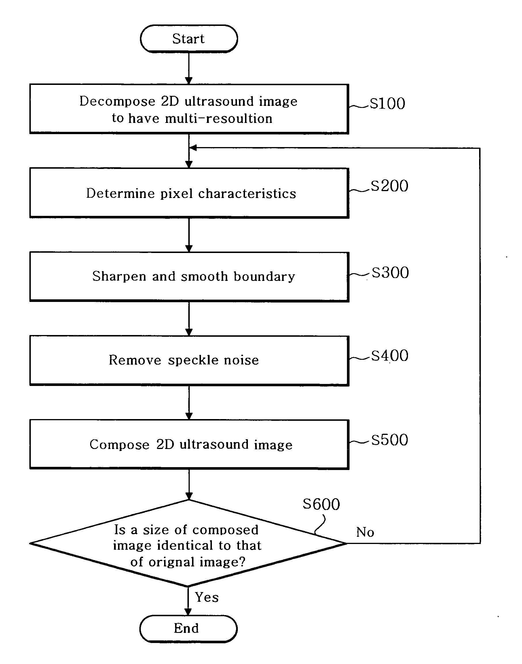
[0011]FIG. 1 is a flow chart showing a process for enhancing an image quality of a 2-dimensional (2D) ultrasound image through a post-processing in accordance with the present invention.

[0012] Referring to FIG. 1, an ultrasound image is decomposed to obtain a multi-resolution at step S100. The ultrasound image may be an image before or after performing a scan conversion process. The multi-resolution analysis is carried out by decomposing an arbitrary image into a plurality of images having a multi-resolution and analyzing the decomposed images. A high frequency component and a low frequency component in the ultrasound image can be obtained through the multi-resolution analysis. In order to obtain the image having the multi-resolution, the wavelet transform and Laplacian pyramid code may preferably be used. In accordance with the present invention, any image decomposition method can be applied to obtain the image having the multi-resolution. Further, the decomposition process can be...


PUM


Abstract

The present invention relates a method and an apparatus for enhancing ultrasound image quality through a post-processing. A method for enhancing an image quality of a two-dimensional (2D) ultrasound image, comprises the steps of: a) decomposing the 2D ultrasound image into a plurality of images having a multi-resolution by N levels, wherein N is a positive integer; b) determining characteristics of each pixel in the decomposed image; c) performing an enhancement process for the decomposed image based on the pixel characteristics; d) performing 1st-level composition for the decomposed image; and e) repeatedly performing steps b) to d) until a size of the composed image is identical to that of the 2D ultrasound image.

Description

FIELD OF THE INVENTION [0001] The present invention generally relates to an ultrasound diagnostic device, and more particularly to a method and an apparatus for enhancing the image quality of an ultrasound image in an ultrasound diagnostic device. BACKGROUND OF THE INVENTION [0002] An ultrasound diagnostic device is used in various medical fields. The ultrasound diagnostic device is adapted to transmit ultrasound signals to organs and acquire an ultrasound image by detecting the ultrasound signals that reflect from the organs. When the ultrasound signals are transmitted to the organs, the reflection and scattering of the ultrasound signals occur at the same time, thus generating a speckle noise. The speckle noise degrades not only the ultrasound image quality but also the accuracy of displaying important parts such as the boundary between a target object and a background. Also, the speckle noise makes it difficult to analyze the ultrasound image and to distinguish different types of...

Claims


Application Information

Patent Timeline
IPC IPC(8): A61B8/00
CPCG06T5/002G06T5/003G06T2207/10132G06T2207/20016G06T2207/30004A61B8/00
Inventor KIM, CHEOL ANSONG, YOUNG SEUKYANG, EUN HORA, JONG BEOMKIM, YONG SUNYU, DONG HOON
Owner KOREA ADVANCED INST OF SCI & TECH
Why Eureka
Try Eureka