
Early prostate cancer antigens (epca), polynucleotide sequences encoding them, and their use

a prostate cancer and antigen technology, applied in the field of early detection of prostate cancer, can solve the problems of false negative biopsy reports and absence of epca in the prostate, and achieve the effect of limiting the number of biopsies in an individual and early detection

Inactive Publication Date: 2006-07-06
UNIVERSITY OF PITTSBURGH +1
View PDF16 Cites 0 Cited by
  • Summary
  • Abstract
  • Description
  • Claims
  • Application Information

AI Technical Summary

Benefits of technology

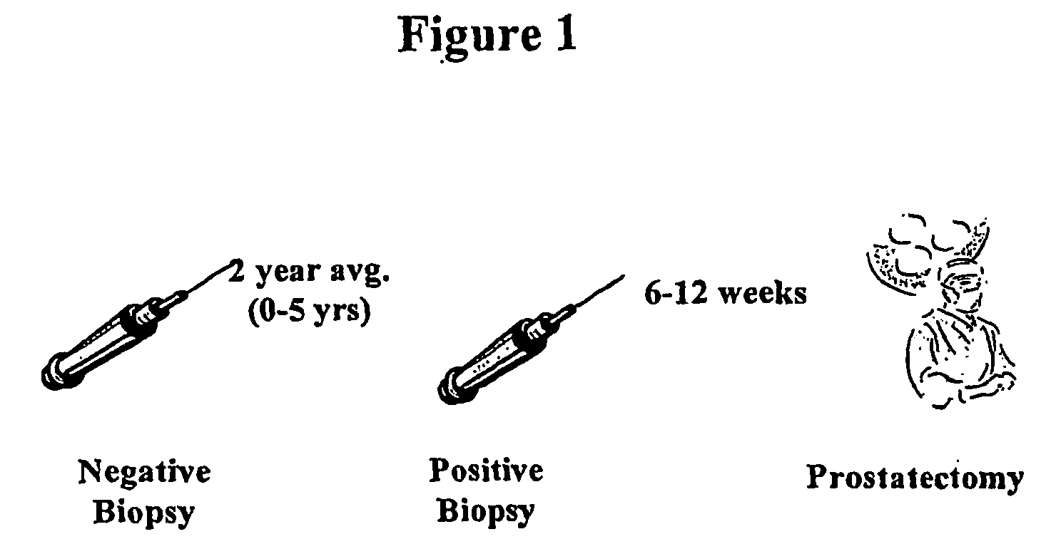
[0020] A novel human protein, designated herein as “early prostate cancer antigen (EPCA),” is expressed throughout the prostate of individuals with prostate cancer and is clinically useful for early detection of individuals with the disease, even in biopsied tissue samples that are morphologically normal. Indeed, the EPCA marker can detect the disease in individuals more than two years prior to morphological manifestation. Thus, anti-EPCA antibodies are useful as an adjunct to pathological examination of prostatic biopsies to detect prostate cancer earlier than using repeated biopsies as well as potentially limiting the number of biopsies in an individual.
[0022] In another aspect, the present invention, directed to EPCA, can be employed to predict the occurrence of prostate cancer and to monitor the progression of prostate cancer. EPCA is expressed at higher levels in aggressive prostate cancer than in non-aggressive prostate cancer. It stains the intra-prostatic tissue, the lymph nodes and seminal vesicles of individuals with metastatic prostate cancer. EPCA also is present in normal tissue adjacent to tumor tissue of individuals having prostate cancer. However, EPCA is absent in the prostate of individuals without prostate cancer. Thus, detection methods employing EPCA as a prostate cancer biomarker decrease the risk of false negative biopsy reports due to sampling errors, because EPCA is present in both normal and diseased prostate tissues of individuals having prostate cancer.
[0023] According to another aspect, the present invention is directed to EPCA and fragments thereof, which are expressed throughout the prostate of individuals with prostate cancer, even in biopsy samples that are morphologically negative. Surprisingly, EPCA is detectable in the prostate of these individuals at least two years prior to morphological detection of prostate cancer. Antibodies against EPCA or fragments thereof are useful as an adjunct to pathologic examination of prostatic biopsies, to detect prostate cancer earlier, and thus avoid or reduce the need for repeated biopsies.

Problems solved by technology

However, EPCA is absent in the prostate of individuals without prostate cancer.
Thus, detection methods employing EPCA as a prostate cancer biomarker decrease the risk of false negative biopsy reports due to sampling errors, because EPCA is present in both normal and diseased prostate tissues of individuals having prostate cancer.

Method used


Image

  • Early prostate cancer antigens (epca), polynucleotide sequences encoding them, and their use
  • Early prostate cancer antigens (epca), polynucleotide sequences encoding them, and their use
  • Early prostate cancer antigens (epca), polynucleotide sequences encoding them, and their use

Examples


examples

Isolation and Sequencing of Rat Nuclear Matrix Proteins

[0162] A rat model system was utilized to identify targets, which were then investigated in human samples.

[0163] The G, AT2.1 and MLL sublines of the Dunning R3327 rat prostate adenocarcinoma cell line were cultured in RPMI 1640 containing 10% fetal bovine serum, 250 nM dexamethasone, penicillin-G and streptomycin both at 100 units / ml. The cells were then harvested and fractionated to isolate nuclear matrix proteins, as described below.

[0164] The Dunning R3327 AT2.1 rat prostate tumors were transplanted subcutaneously into male Copenhagen rats and harvested when the tumor weights reached 34 grams. Normal rat dorsal prostates were obtained from mature intact male Sprague-Dawley rats (300-350 g) obtained from Charles River (Wilmington, Mass.). Tumor and tissue samples were fractionated to isolate nuclear matrix proteins as described below.

[0165] Normal and tumor prostate tissue samples were obtained from patients undergoing s...


PUM

PropertyMeasurementUnit
weightsaaaaaaaaaa
pHaaaaaaaaaa
pHaaaaaaaaaa

Abstract

A novel prostate cancer marker is described that is found in cancerous and normal prostate cells of individuals that have prostate cancer but is not found in the prostate of individuals without prostate cancer. The marker also is present in normal tissue adjacent to tumor tissue in individuals having prostate cancer. The marker, however, is absent in the prostate of individuals without prostate cancer. Methods employing the novel prostate cancer marker of the invention to predict the occurrence of the prostate disease, monitor the progression of prostate cancer and effect the treatment of prostate cancer, also are described.

Description

CROSS-REFERENCE TO RELATED PATENT APPLICATIONS [0001] This application is a continuation-in-part of U.S. application Ser. No. 10 / 375,058, filed Feb. 28, 2003, which is a continuation-in-part of U.S. application Ser. No. 09 / 418,839, filed Oct. 15, 1999.STATEMENT REGARDING GOVERNMENT SPONSORED RESEARCH [0002] This invention was made with support from the National Cancer Institute under grant No. R29 CA65463-01. Thus, the government may have certain rights to this invention.FIELD OF THE INVENTION [0003] This invention relates generally to compositions and methods for detecting cancer in mammals, including humans. More particularly, the invention relates to the use of a protein, herein designated early prostate cancer antigen (EPCA), for diagnosing and treating prostate cancer, and to methods of assessing the aggressiveness and metastatic potential of prostate cancer by monitoring the level of EPCA or fragments thereof. BACKGROUND OF THE INVENTION [0004] Abnormalities of prostate growth...

Claims


Application Information

Patent Timeline
Patent Type & Authority Applications(United States)
IPC IPC(8): G01N33/574G01N33/53A61KA61K31/7088A61K48/00A61P35/00A61P35/04C07K14/47C07K14/82C07K16/18C07K16/32C12N15/12C12P21/08C12Q1/68G01N33/50
CPCC07K14/47Y10T436/143333G01N33/57434C07K14/4748A61P35/00A61P35/04
Inventor GETZENBERG, ROBERT
Owner UNIVERSITY OF PITTSBURGH
Learn More