Use of computer-aided detection system outputs in clinical practice

a detection system and output technology, applied in healthcare informatics, image enhancement, instruments, etc., can solve problems such as poor image quality, missed detections, and improper patient positioning, and achieve efficient and accurate positioning of exact areas

Inactive Publication Date: 2006-08-03
ICAD INC
View PDF49 Cites 59 Cited by
  • Summary
  • Abstract
  • Description
  • Claims
  • Application Information

AI Technical Summary

Benefits of technology

[0012] The present invention provides for the use of CAD output displays for providing accurate representation of areas to undergo re-imaging procedures Since the CAD output is not used during the initial reading, the radiologist does not mark it until a final determination is reached regarding subsequent procedures. Additionally,

Problems solved by technology

Missed detections may be attributed to several factors including poor image quality, improper patient positioning, inaccurate interpretation, fibroglandular tissue obscuration, subtle nature of radiographic findings, eye fatigue, or oversight.
One difficul

Method used


Image

  • Use of computer-aided detection system outputs in clinical practice
  • Use of computer-aided detection system outputs in clinical practice
  • Use of computer-aided detection system outputs in clinical practice

Examples


Embodiment Construction

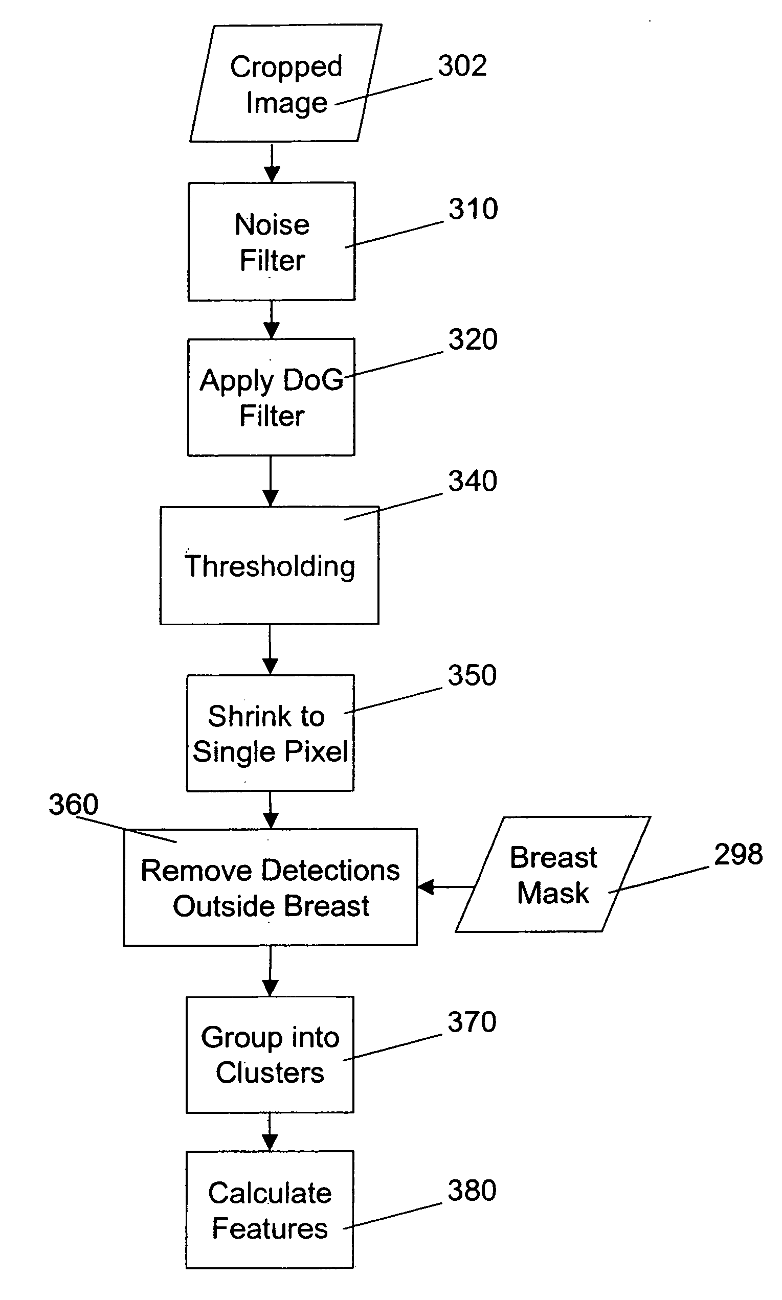
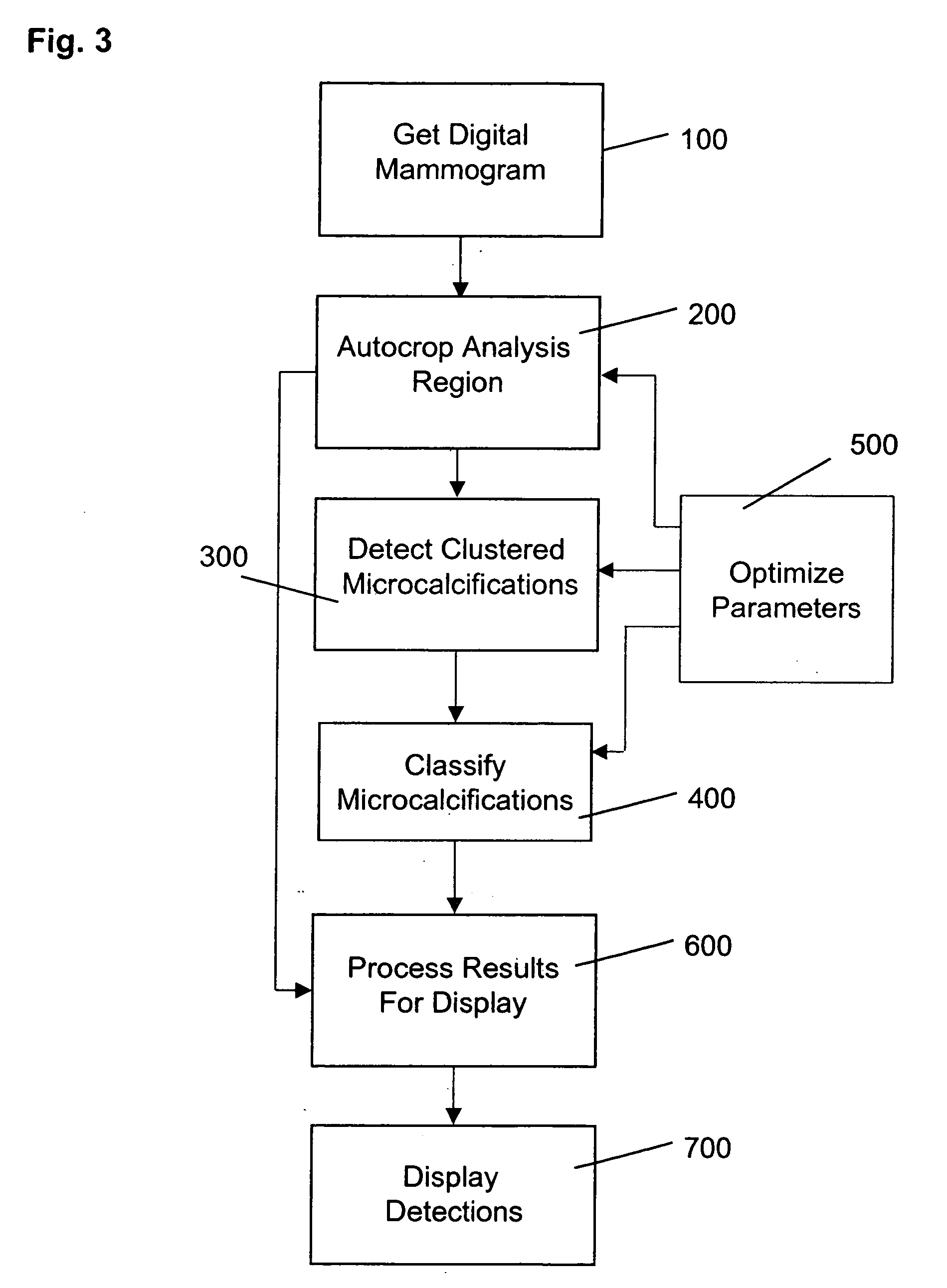
[0054] Referring now to the drawings, wherein like reference numerals designate identical or corresponding parts throughout the several views, and more particularly to FIG. 3 thereof, there is shown a flow diagram illustrating a sequence of steps performed in order to detect the locations of clusters of microcalcifications within a digital mammogram.

[0055] In a first step 100, a digital mammogram is obtained using hardware such as digital mammography systems, or by digitizing mammography films using laser or charge-coupled device (CCD) digitizers. In an optimized cropping step 200, a rectangular analysis region containing breast tissue is segmented from the digital mammogram image and a binary mask corresponding to the breast tissue is created for use in later processing steps to decrease the time required for processing the mammogram image. The binary mask is also used to limit detections to areas of the image containing breast tissue

[0056] Clustered microcalcifications are detec...


PUM


Abstract

The present invention provides for the use of computer-aided detection (CAD) system output displays for providing accurate representations of areas for subsequent exams. Since the CAD output, unlike the original medical imagery, is not used during the initial reading, the radiologist does not mark it until a final determination is reached regarding subsequent procedures. Additionally, since the CAD output contains versions of the original imagery, the regions indicated by the radiologist are shown in the context of the particular anatomical detail for a given patient. This detail assists the technologist, other physicians and patients in more efficiently and accurately locating the exact area for subsequent exams.

Description

CROSS-REFERENCE TO RELATED APPLICATIONS [0001] This application is a continuation-in-part of U.S. application Ser. No. 10 / 674,232, filed on Sep. 29, 2003, now allowed, which is a continuation-in-part of U.S. application Ser. No. 10 / 280,237, filed Oct. 25, 2002, now U.S. Pat. No. 6,650,766, which is a continuation of U.S. application Ser. No. 09 / 938,908, filed Aug. 24, 2001, now U.S. Pat. No. 6,556,699, which is a continuation of U.S. application Ser. No. 09 / 758,889, filed Jan. 11, 2001, now U.S. Pat. No. 6,389,157, which is a continuation of U.S. application Ser. No. 09 / 416,437, filed Oct. 12, 1999, now U.S. Pat. No. 6,205,236, which is a continuation of U.S. application Ser. No. 09 / 14 1,802, filed Aug. 28, 1998, now U.S. Pat. No. 5,999,639, which claims the benefit of U.S. Provisional Application No. 60 / 057,801, filed Aug. 28, 1997, U.S. Provisional Application No. 60 / 066,996, filed Nov. 28, 1997, and U.S. Provisional Application No. 60 / 076,760, filed Mar. 3, 1998, the entire discl...

Claims


Application Information

Patent Timeline
IPC IPC(8): G06K9/00G06V10/25G06V10/28G16H30/20G16H30/40
CPCG06F19/321G06F19/345G06K9/3233G06K9/38G06K9/622G06T7/0012G06T7/0081G06T7/0091G06T2207/10116G06T2207/20084G06T2207/20148G06T2207/30068Y10S128/922G06T7/11G06T7/155G06T7/136G16H50/20G16H30/20G16H30/40G06V10/25G06V10/28G06F18/232
Inventor ROGERS, STEVEN K.
Owner ICAD INC
Why Eureka
Try Eureka