Circular ultrasound tomography scanner and method

Inactive Publication Date: 2006-08-03
HELIX MEDICAL SYST
View PDF9 Cites 42 Cited by
  • Summary
  • Abstract
  • Description
  • Claims
  • Application Information

AI Technical Summary

Benefits of technology

[0013] at least one drive unit attached to the housing and to the at least one sc

Problems solved by technology

All these methods have shortcomings related to the flaccid nature of the breast which leads to difficulty in manipulating and orienting the organ.
Additionally, some of these methods are very uncomfortable for the patient, the pain often associated with the forceful compression of the breast between plates in mammography being a prime example.
Consequently, during use there is a tendency for the breast to move together with the rotating housing.
Such movement is minimized by introducing

Method used


Image

  • Circular ultrasound tomography scanner and method
  • Circular ultrasound tomography scanner and method
  • Circular ultrasound tomography scanner and method

Examples


Embodiment Construction

[0041] In the following description, components having similar functionality in more than one figure will be referenced in all figures by identical reference numerals.

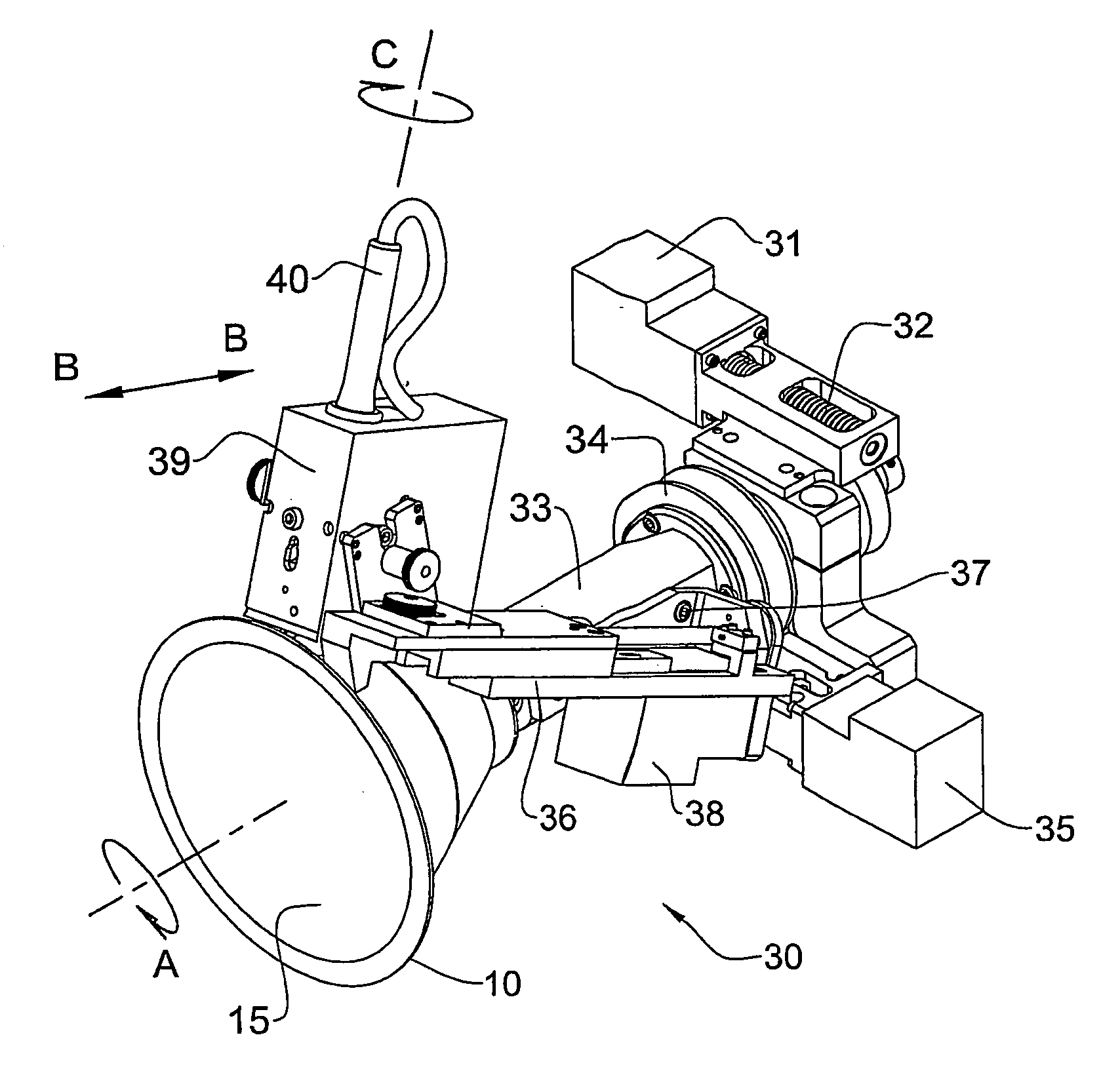
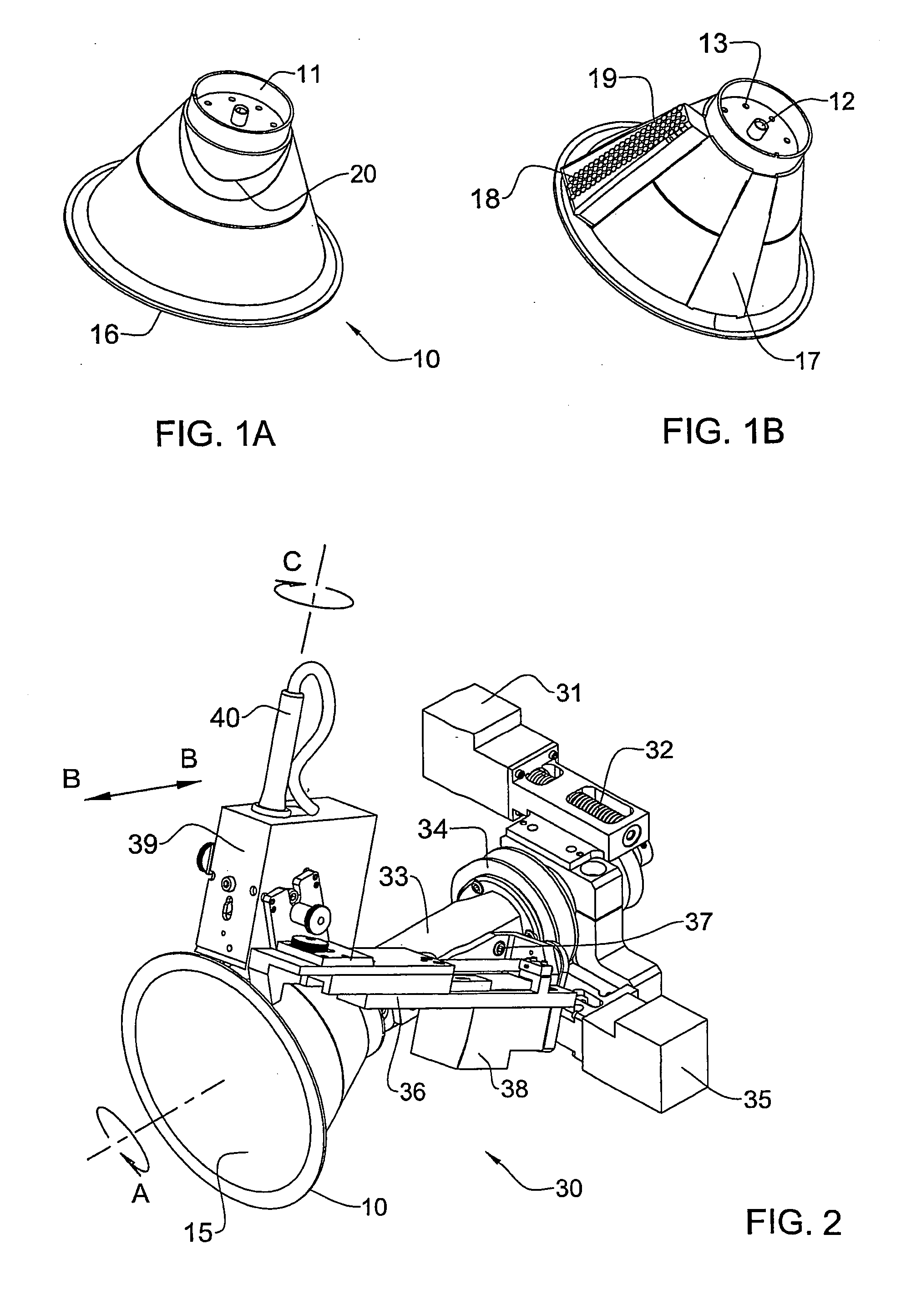
[0042]FIGS. 1a and 1b show pictorially a substantially conical housing 10 according to an exemplary embodiment of the invention for accommodating a breast (not shown) during scanning. The housing 10, which may be disposable, has a first end 11 having a threaded portion 12 for threadably attaching to a motor unit so as to allow rotation of the housing. Apertures 13 formed in the first end 11 of the housing cooperate with a vacuum pump (not shown) for allowing suction to be applied. The apertures 13 thus operate as a securing unit for securing the breast within the housing and thereby avoid dislocation thereof during scanning, which if not prevented would derogate from the accuracy of subsequent measurements. A second end 15 of the housing is open to allow for insertion of the breast and is provided with a circumferenti...


PUM


Abstract

A portable mechanical high-precision device (30) for performing circular or helical scanning of a patient's organ (71) or body surface for tissue diagnosis and/or treatment includes a substantially hollow housing (10) for accommodating the organ therein and a securing unit (61) for securing the housing to the organ or body surface during scanning so that the organ or body surface is substantially fixed relative to the housing. At least one drive unit (31, 35, 38) is attached to the housing and to at least one scan head (39) for allowing unlimited rotation of the scan head relative to the housing.

Description

FIELD OF THE INVENTION [0001] This invention relates to ultrasound tomography particularly, albeit not exclusively, for diagnosis and treatment of breast tumors. BACKGROUND OF THE INVENTION [0002] It is known to obtain ultrasound images of the breast using a probe placed against the breast. Stereotactic biopsy techniques are done in a similar fashion; with a patient lying prone, with the breast hanging through a hole in the table. All these methods have shortcomings related to the flaccid nature of the breast which leads to difficulty in manipulating and orienting the organ. Additionally, some of these methods are very uncomfortable for the patient, the pain often associated with the forceful compression of the breast between plates in mammography being a prime example. [0003] US 2003 / 0233110A1 (Jesseph) published Dec. 18, 2003 and entitled “Device and method for improved diagnosis and treatment of cancer” discloses a fixation device, which fixes a breast in distended, stable positi...

Claims


Application Information

Patent Timeline
IPC IPC(8): A61B8/00A61B8/06A61B8/08A61B8/14A61B19/00
CPCA61B8/06A61B8/0825A61B8/13A61B8/14A61B8/4461A61B8/488A61B2019/205A61B90/17
Inventor AMARA, ARIEAMARA, AVI
Owner HELIX MEDICAL SYST
Why Eureka
Try Eureka