Method and device for canulation and occlusion of uterine arteries

a technology of uterine arteries and cannulation, which is applied in the field of methods and devices for cannulation and occlusion of uterine arteries, can solve the problem that methods require a relatively high level of catheterization skill

Inactive Publication Date: 2006-08-10
BOSTON SCI SCIMED INC
View PDF8 Cites 27 Cited by
  • Summary
  • Abstract
  • Description
  • Claims
  • Application Information

AI Technical Summary

Benefits of technology

[0004] The present invention is further directed to a device for treating uterine fibroids comprising an elongated sheath sized for insertion into a uterine artery via an incision in the vaginal fornix, the sheath including a sheath lumen extending from a first sheath opening formed in a proximal end of the sheath to a second sheath opening formed in a distal end of the sheath and a body a distal end of which is connected to the proximal end of the sheath, the body including a body lumen extending therethrough from a first body opening at a proximal end of the body and a second body opening at the distal end thereof, the second body lumen communicating with the sheath lumen in combination with a hemostatic valve controlling the flow of blood through the body lumen.

Problems solved by technology

However, these methods require a relatively high level of catheterization skill and have, therefore, been primarily performed by interventional radiologists in catheterization labs or angiography suites.

Method used


Image

  • Method and device for canulation and occlusion of uterine arteries
  • Method and device for canulation and occlusion of uterine arteries
  • Method and device for canulation and occlusion of uterine arteries

Examples


Embodiment Construction

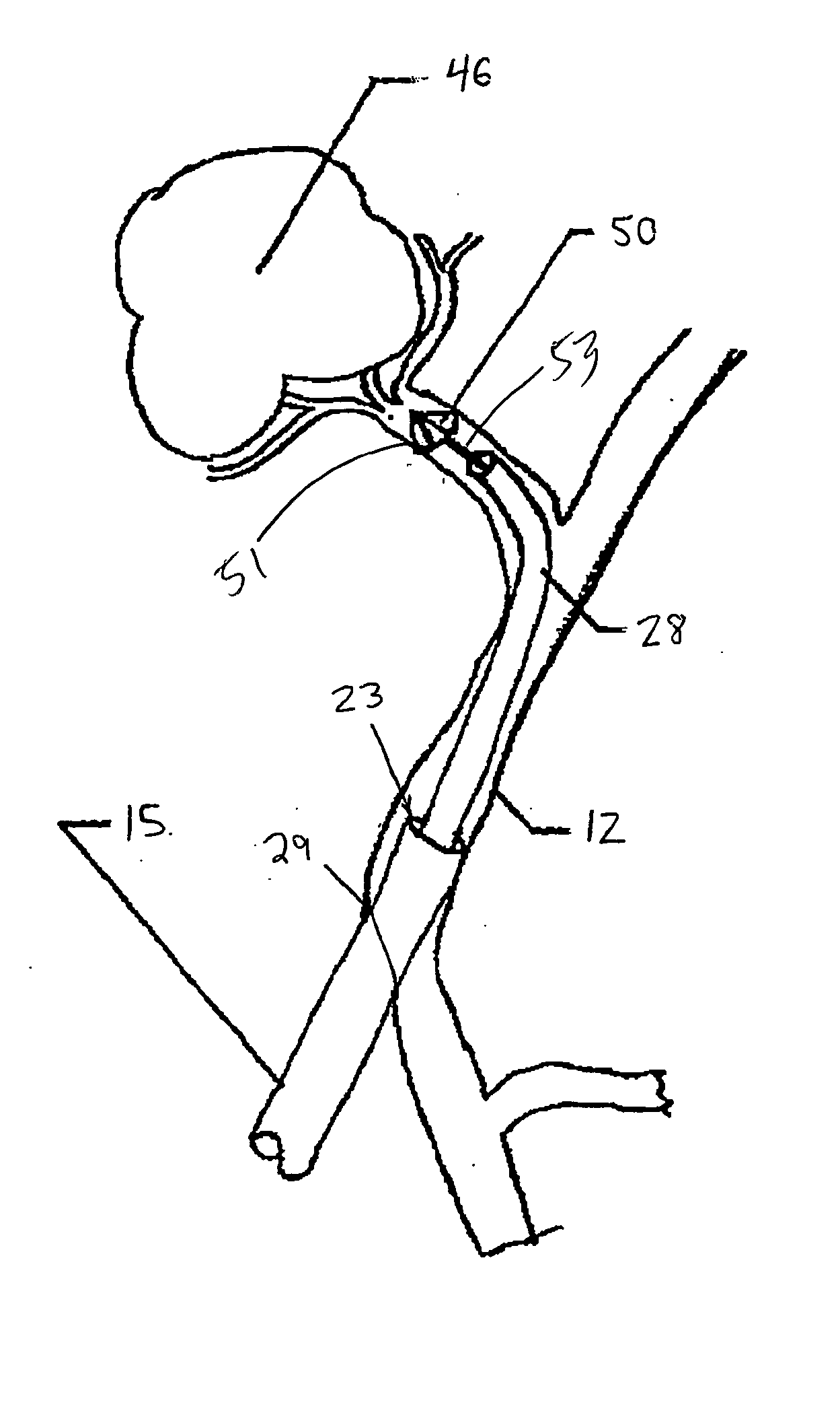
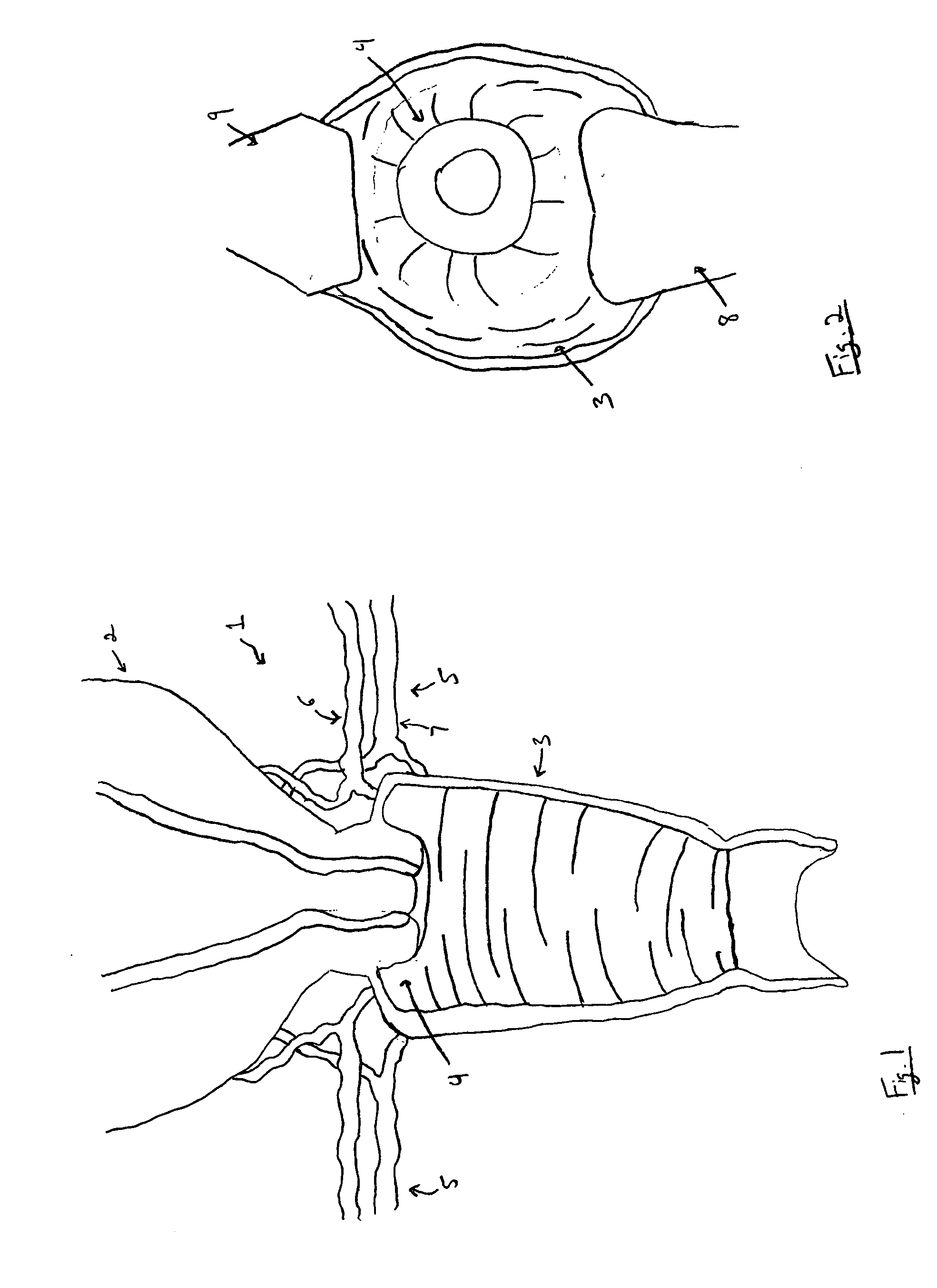
[0018] The present invention is directed to a simplified method and device for treating fibroids without requiring the same high level of catheterization skills required for the prior methods. FIG. 1 shows the structure of the female reproductive system generally seen at 1. The uterus 2 is superior to the vagina 3, with the cervix 4 comprising a lowermost portion of the uterus 2, which connects to the vagina 3. A lowermost portion of the cervix 4 is exposed to the vagina 3. The uterine blood vessels 5, located superior to the vagina 3 and inferior to the uterus 2, include a series of uterine arteries 6 and a series of uterine veins 7.

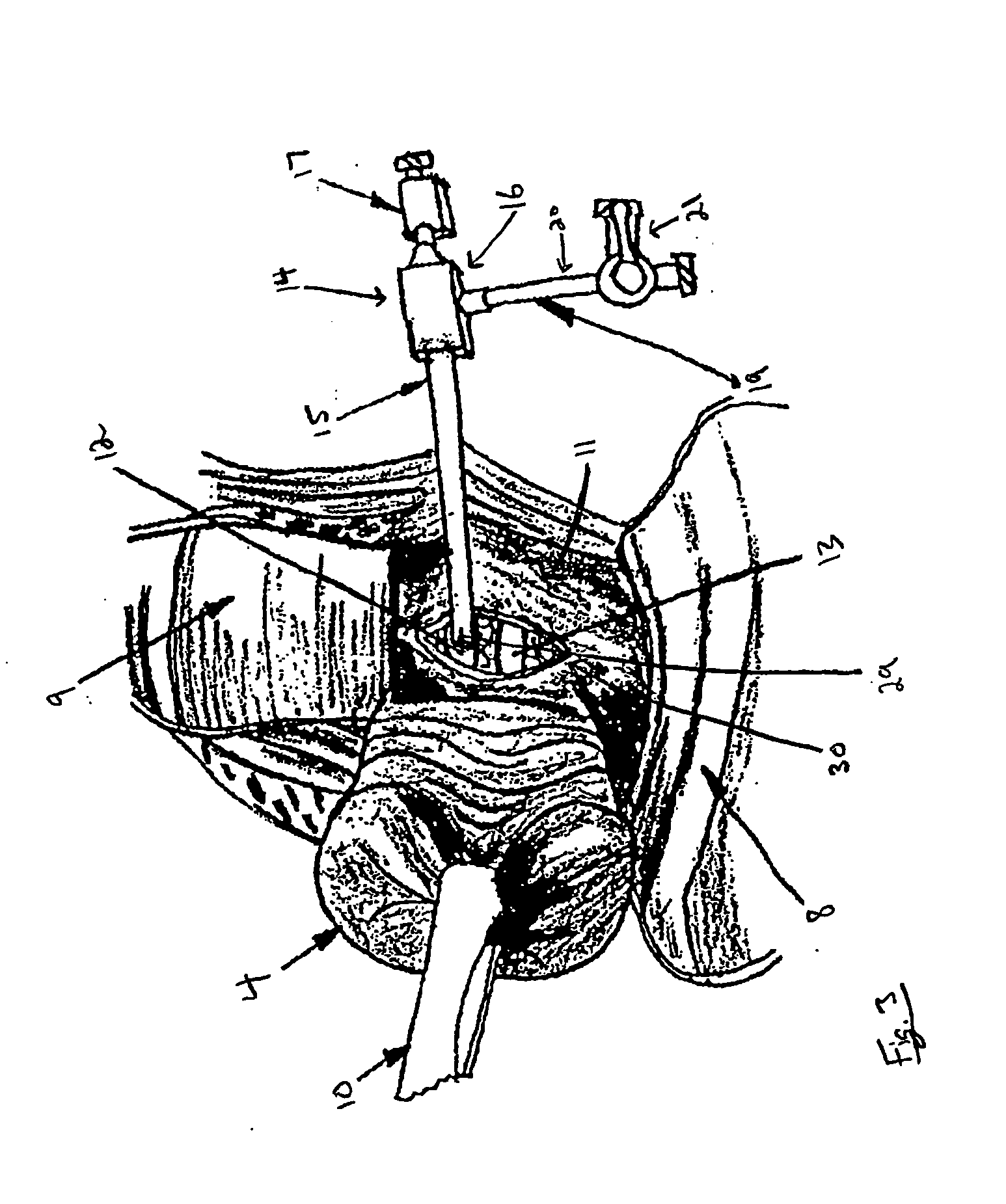
[0019] In accordance with the present method, FIG. 1 depicts an initial view of the uterine blood vessels 5. As seen in FIG. 2, visualization of the cervix 4 may be maximized by inserting a speculum 8 into the vagina 3 at a lower position while using a retractor 9 at an upper position of the vagina 3. As would be understood by those of skill in the art...


PUM


Abstract

A method for treating a uterine fibroid comprises forming an incision in a vaginal fornix to expose a first blood vessel supplying the fibroid, forming an opening in the first blood vessel and inserting an introducer into the first blood vessel via the opening in combination with the steps of advancing a catheter to a desired position within the first blood vessel via the introducer and introducing an occlusive agent into the first blood vessel through the catheter to block blood flow through the first blood vessel. A device for treating uterine fibroids comprises an elongated sheath sized for insertion into uterine arteries via an incision in the vaginal fornix, the sheath including a sheath lumen extending from a first sheath opening formed in a proximal end of the sheath to a second sheath opening formed in a distal end of the sheath and a body a distal end of which is connected to the proximal end of the sheath, the body including a body lumen extending therethrough from a first body opening at a proximal end of the body and a second body opening at the distal end thereof, the second body lumen communicating with the sheath lumen in combination with a hemostatic valve controlling the flow of blood through the body lumen.

Description

FIELD OF INVENTION [0001] The present invention relates generally to methods and devices for canulation and occlusion of uterine arteries for the treatment of fibroids. BACKGROUND [0002] Several procedures are available for the treatment of uterine leiomyoma (fibroids) including the delivery of occlusive agents to the uterine arteries to prevent blood flow to the fibroids via catheters introduced into the uterine arteries via access from the femoral artery. However, these methods require a relatively high level of catheterization skill and have, therefore, been primarily performed by interventional radiologists in catheterization labs or angiography suites. SUMMARY OF THE INVENTION [0003] The present invention is directed to a method for treating a uterine fibroid comprising forming an incision in a vaginal fornix to expose a first blood vessel supplying the fibroid, forming an opening in the first blood vessel and inserting an introducer into the first blood vessel via the opening ...

Claims


Application Information

Patent Timeline
Patent Type & Authority Applications(United States)
IPC IPC(8): A61M29/00
CPCA61B17/02A61B17/12022A61B17/12131A61B17/12159A61B17/12172A61B17/12186A61B17/3415A61B17/42A61B2017/1205A61M39/06A61B2017/12054
Inventor MCINTYRE, JON T.MADDEN, MICHAEL
Owner BOSTON SCI SCIMED INC
Why Eureka
Try Eureka