Method and apparatus to visualize the coronary arteries using ultrasound

a technology of coronary arteries and ultrasound, applied in the field of medical ultrasound, can solve the problems of arteries that do not permit them to be seen for any length, and it takes great skill and knowledge to recognize arteries, so as to reduce noise, minimize unwanted angulation, and improve three-dimensional images

Active Publication Date: 2007-10-11
MAUI IMAGING
View PDF85 Cites 140 Cited by
  • Summary
  • Abstract
  • Description
  • Claims
  • Application Information

AI Technical Summary

Benefits of technology

[0025]It is still another object of the present invention to provide improved three dimensional images of coronary arteries by utilizing linear filtering to reduce noise, using nonlinear techniques, such as neural networks, to highlight arteries and veins in contrast to other tissue and the heart chambers, storing numerous processed 2D slices into a 3D volume, and then projecting the volume with varying view angles controlled by a pointing device.
[0026]An even further object of the present invention is to provide gyroscopic stabilization to the ultrasound probe to minimize unwanted angulation during data collection.

Problems solved by technology

However, as Feigenbaum states, the curved nature of the arteries usually does not permit them to be seen for any length in an individual frame.
In addition, it takes great skill and knowledge to recognize the arteries when they do come into view.

Method used


Image

  • Method and apparatus to visualize the coronary arteries using ultrasound
  • Method and apparatus to visualize the coronary arteries using ultrasound
  • Method and apparatus to visualize the coronary arteries using ultrasound

Examples


Embodiment Construction

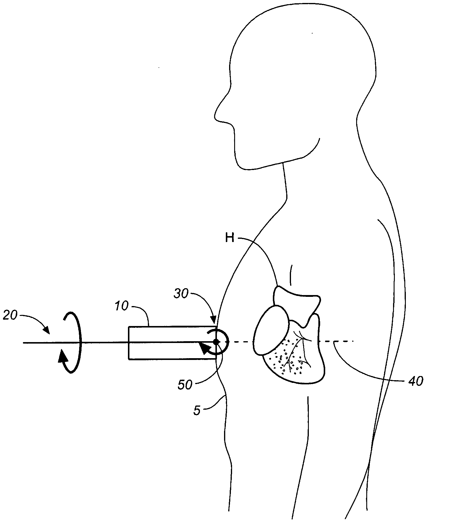
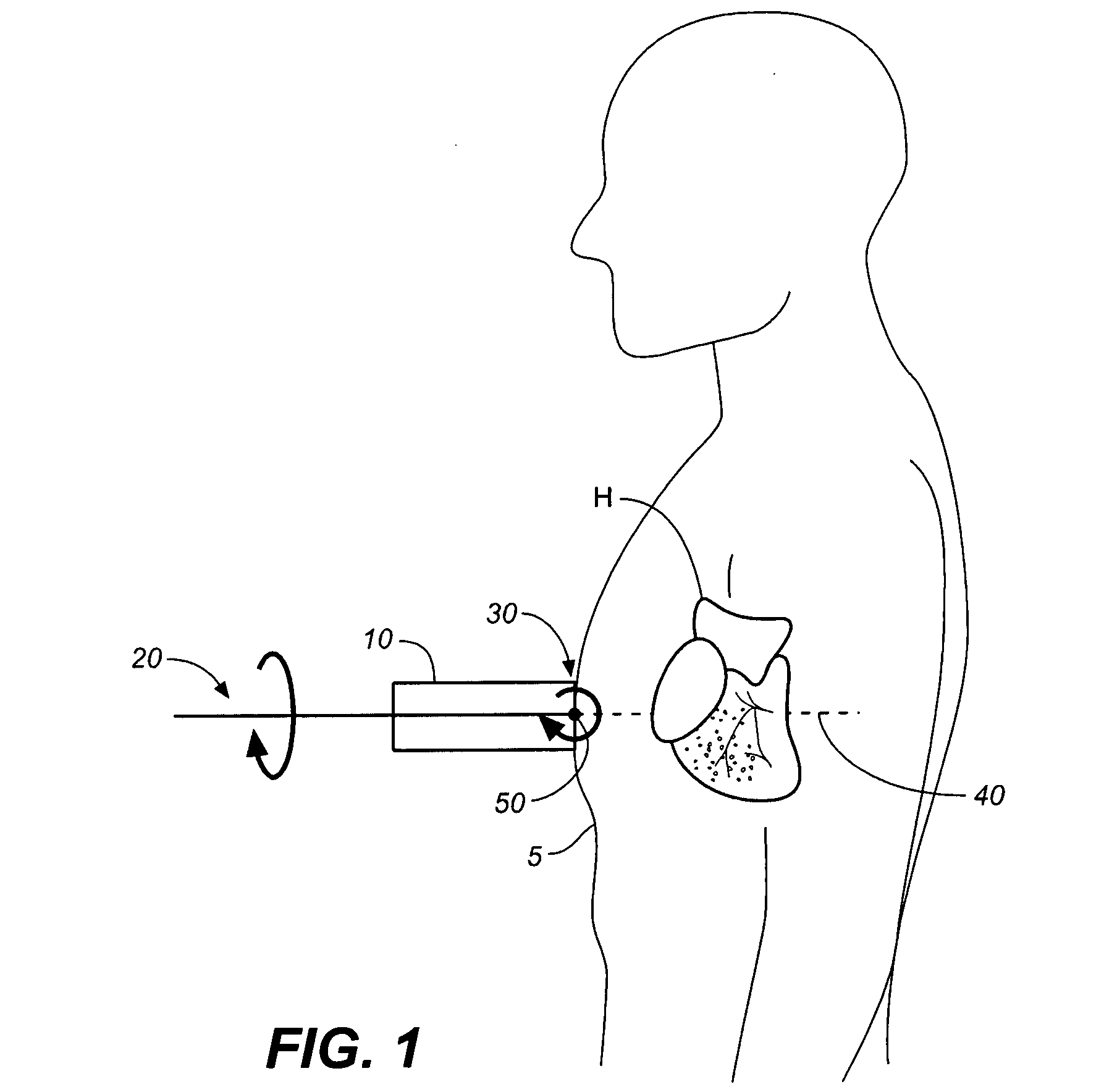
[0036]The present invention is a method and apparatus that renders a projection of images of the coronary arteries in three dimensions using ultrasound. In its most essential aspect, this is accomplished by first producing a 3D array of voxels indicating the blood-filled areas of the heart. Next, a 2D image of the blood-filled areas is projected as a function of view angle and rotation, and this allows an observation and evaluation of the blood-filled areas from a number of view angles such that the coronary arteries and veins are seen unobscured by the major chambers of the heart. The objective is to provide a non-invasive screening test to assess the patency of the coronary arteries. It is hoped that in addition to detecting complete blockages of the arteries, it will also be possible to assess the degree of obstruction in partially occluded arteries.

[0037]Several methods are available to obtain the necessary three dimensional information using ultrasound. Two methods have been pu...


PUM


Abstract

A non-invasive screening technique for visualizing coronary arteries which overcomes the problems of visualizing the curved arteries by projecting the three dimensional volume of the arteries onto a two dimensional screen. Blood filled areas, and in particular, the coronary arteries and veins, are highlighted to contrast them with other nearby tissues using non-linear classification and segmentation techniques. Data is gathered as a sequence of 2D slices stored as a 3D volume. Software is employed to interpolate voxels intermediate to the slices. Wiener filtering or LMS spatial filtering can be implemented on each 2D scan to improve lateral resolution and reduce noise prior to the use of the scan data with the classification and segmentation algorithms. A traditional handheld ultrasound probe is employed to enable the technician to locate the area of interest, but a gyroscopic stabilizer is added to minimize unwanted variation on two axes of rotation while scanning through angles on the third axis of rotation.

Description

CROSS REFERENCES TO RELATED APPLICATIONS[0001]The present application claims the benefit of U.S. Provisional Patent Application Ser. No. 60 / 765,887, filed Feb. 6, 2006, (Feb. 6, 2006).STATEMENT REGARDING FEDERALLY SPONSORED RESEARCH OR DEVELOPMENT[0002]Not applicable.THE NAMES OR PARTIES TO A JOINT RESEARCH AGREEMENT[0003]Not applicable.INCORPORATION-BY-REFERENCE OF MATERIAL SUBMITTED ON A COMPACT DISC[0004]Not applicable.BACKGROUND OF THE INVENTION[0005]1. Field of the Invention[0006]The present invention is generally related to medical ultrasound, and more particularly to a non-invasive screening method and apparatus for visualizing coronary arteries which solves artery curvature problems by projecting the three dimensional volume onto a two dimensional screen, highlighting the arteries and veins in contrast to other tissue and the heart chambers by using nonlinear techniques, storing many processed 2D slices into a 3D volume, and projecting the volume with varying view angles con...

Claims


Application Information

Patent Timeline
Patent Type & Authority Applications(United States)
IPC IPC(8): A61B8/00
CPCA61B5/02007G06T2207/30101A61B5/7264A61B8/06A61B8/08A61B8/0883A61B8/0891A61B8/4254A61B8/483A61B8/543G06T7/0012G06T2207/10136G06T2207/20092G06T2207/30048A61B5/7257
Inventor SPECHT, DONALD F.
Owner MAUI IMAGING
Why Eureka
Try Eureka