Methods and tools for accessing an anatomic space

an anatomic space and access method technology, applied in the field of medical devices and methods, can solve the problems of unattractive scars, carries additional complications, and traumatized thoracic muscles and ribs, and achieves the effect of more robust fixation

Active Publication Date: 2006-09-07
MEDTRONIC INC
View PDF26 Cites 168 Cited by
  • Summary
  • Abstract
  • Description
  • Claims
  • Application Information

AI Technical Summary

Benefits of technology

[0020] The suction pads advantageously facilitated cutting into or through the outer tissue layer with a cutting instrument. Initially, suction is applied so that the suction pads grasp the surface of the outer tissue layer. A cutting instrument or tool can then be introduced through the working lumen to dispose a cutting element at the tissue surface. The suction pads are also spread apart so that the tissue surface is tensioned during cutting. In this way, the perforation or hole that is cut is widened by the applied tension. The widened hole can then be maintained for passage of further devices into or through the hole.
[0025] Due to their redundancy, the suction ports of the suction pads provide more robust fixation to the outer tissue layer than a single large area suction port. At least some of the suction ports readily engage the tissue surface under low suction force to enable lifting of the outer tissue layer or tracking movement of the outer tissue layer. Engagement of tissue surface areas by all of the suction ports is not necessary. Similarly, the loss of engagement of some of suction ports with the tissue surface areas does not result in complete loss of engagement as is the case when an edge of a single large suction port releases from the tissue surface of the outer tissue layer.
[0026] A cutting instrument, e.g., a knife, a needle, a stiff guidewire tip, an electrosurgical cutting tool, surgical scissors or other piercing or cutting tool, can then be introduced through the working lumen to perforate the outer tissue layer and form an access hole or perforation there through creating access into the anatomic space while the access tube stabilizes the outer tissue layer. Advantageously, there is no suction applied through the working lumen that is necessary to maintain the attachment to the outer tissue layer while it is being perforated or other instruments are advanced through the perforation. The fixation of the outer tissue layer is not lost when the outer tissue layer is perforated. Moreover, it is simpler to advance instruments through the working lumen from a proximal lumen end opening that is exposed to the atmosphere.
[0028] The tubular access sleeve can be circular or oval or have any other desirable cross-section shape. The tubular access sleeve can be straight, curved or formed with a bend or formed of a bendable material to be shaped by the user. The tubular access sleeve and the suction tool are formed of non-conductive materials to minimize electrical hazards. The tubular access sleeve and the suction tool can be provided in a range of diameters and lengths to function as described herein to access any desired site in the body of adult and pediatric patients.

Problems solved by technology

As a result, these operations typically require large numbers of sutures or staples to close the incision and 5 to 10 wire hooks to keep the severed sternum together.
Such surgery often carries additional complications such as instability of the sternum, post-operative bleeding, and mediastinal infection.
The thoracic muscle and ribs are also severely traumatized, and the healing process results in an unattractive scar.
Therefore, much effort has been expended to develop medical devices and techniques to access the pericardial space employing such minimally invasive percutaneous procedures.
One difficulty has been that normally the pericardial space is so small or thin that it is difficult to penetrate the pericardium using miniaturized instruments capable of being introduced through a port to the site without also puncturing the underling epicardium and thereby, damaging the myocardium or a coronary vessel.
Proliferative adhesions occur between the pericardium and the epicardium in diseased hearts and hamper access to the pericardial space employing such minimally invasive percutaneous procedures.
It is difficult to introduce and use the forceps through the narrow lumen of a port or sleeve, particularly if the pericardial fluid is under pressure that makes the pericardium taut like an inflated balloon.
Although theoretically plausible, the ability to reliably maintain a vacuum seal against the pericardium when such treatment devices are advanced can be problematic.

Method used


Image

  • Methods and tools for accessing an anatomic space
  • Methods and tools for accessing an anatomic space
  • Methods and tools for accessing an anatomic space

Examples


Embodiment Construction

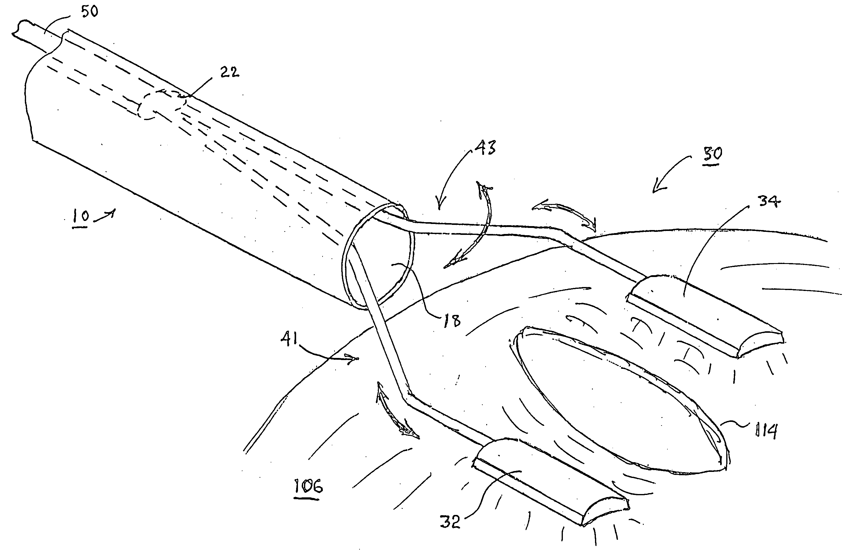
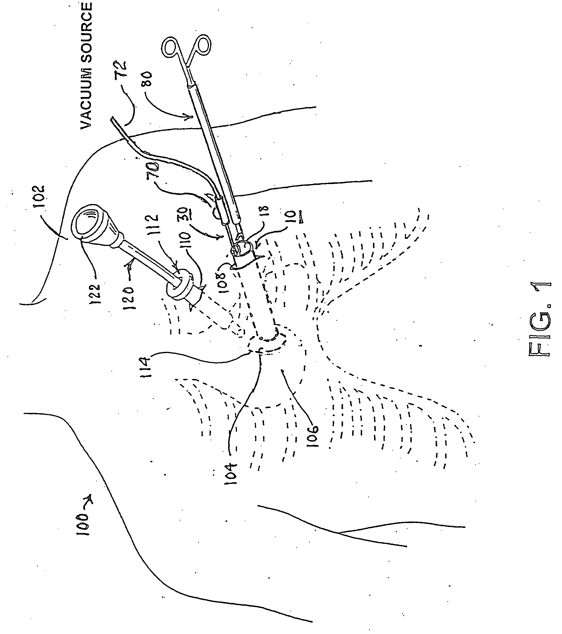
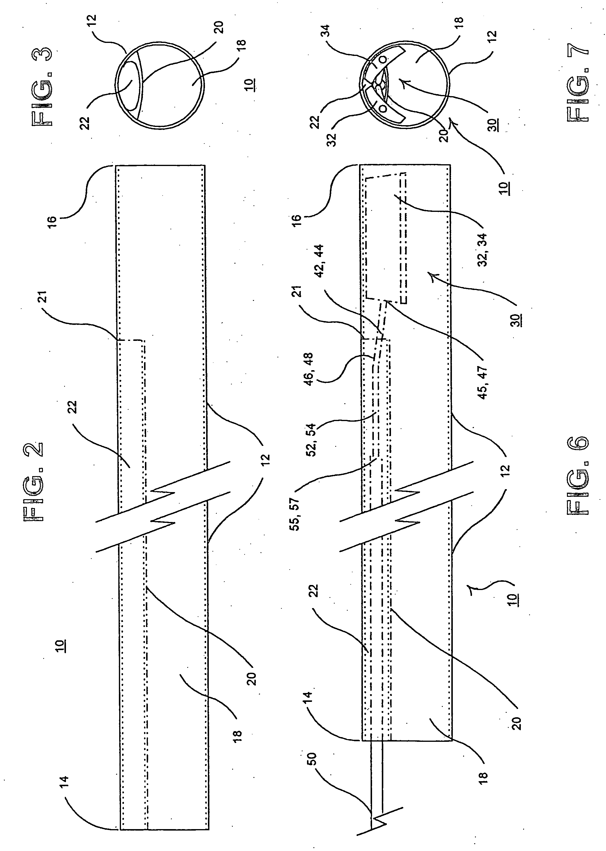
[0039] In the following detailed description, references are made to illustrative embodiments of methods and apparatus for carrying out the invention. It is understood that other embodiments can be utilized without departing from the scope of the invention. Preferred methods and apparatus are described for accessing the pericardial space between the epicardium and the pericardium as an example of accessing an anatomic space between an outer tissue layer and an inner tissue layer.

[0040] The access to the pericardial space in accordance with the present invention facilitates the performance of a number of ancillary procedures. For example, the procedures include introducing and locating the distal end of a catheter or guidewire or an electrode of a cardiac ablation catheter or a pacing lead or a cardioversion / defibrillation lead within the pericardial space and attached to the epicardium or myocardium. Other possible procedures include performing a coronary artery anastomosis in a th...


PUM


Abstract

A tubular access sleeve and suction tool for accessing an anatomic surface or anatomic space and particularly the pericardium to access pericardial space and the epicardial surface of the heart in a minimally invasive manner are disclosed. A suction tool trunk extending through a suction tool lumen of the sleeve is coupled to suction pads at the ends elongated support arms. The suction pads can be retracted into a sleeve working lumen during advancement of the access sleeve through a passage and deployed from the tubular access sleeve lumen and disposed against the an outer tissue layer. Suction can be applied through suction tool lumens to suction ports of the suction pads that fix to the outer tissue layer so as to tension the outer tissue layer and / or pull the outer tissue layer away from an inner tissue layer so that the anatomic space can be accessed by instruments introduced through the working lumen to penetrate the outer tissue layer.

Description

CROSS-REFERENCE TO RELATED APPLICATIONS [0001] This application is a continuation of U.S. Ser. No. 10 / 342,960, filed Jan. 15, 2003.FIELD OF THE INVENTION [0002] The present invention pertains to medical devices and methods for accessing an anatomic surface, muscle layer, vessel or anatomic space of the body and particularly for entering the pericardium to access pericardial space and the epicardial surface of the heart in a minimally invasive manner. BACKGROUND OF THE INVENTION [0003] The human heart wall consists of an inner layer of simple squamous epithelium, referred to as the endocardium, overlying a variably thick heart muscle or myocardium and is enveloped within a multi-layer tissue structure referred to as the pericardium. The innermost layer of the pericardium, referred to as the visceral pericardium or epicardium, clothes the myocardium. The epicardium reflects outward at the origin of the aortic arch to form an outer tissue layer, referred to as the parietal pericardium,...

Claims


Application Information

Patent Timeline
Patent Type & Authority Applications(United States)
IPC IPC(8): A61B1/32A61B17/00A61B17/02A61B17/30
CPCA61B17/0218A61B2017/00247A61B2017/306A61B2018/00392
Inventor GUENST, GARY W.
Owner MEDTRONIC INC
Why Eureka
Try Eureka