
Apparatus and method for non-invasive measurement of intracranial pressure

a non-invasive and intracranial pressure technology, applied in the field of intracranial pressure determination in patients, can solve the problems of inability to adapt to medical practice, limited method, and inability to measure intracranial pressure, and achieve the effect of reducing the amount of blood in the vessel

Inactive Publication Date: 2006-09-14
BRAXTON ERNEST E
View PDF16 Cites 102 Cited by
  • Summary
  • Abstract
  • Description
  • Claims
  • Application Information

AI Technical Summary

Benefits of technology

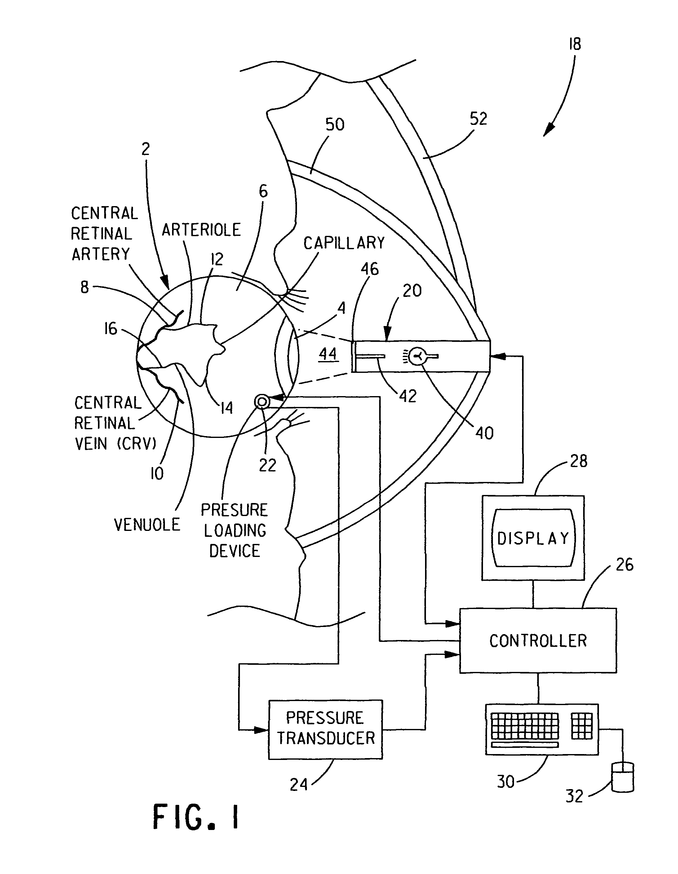
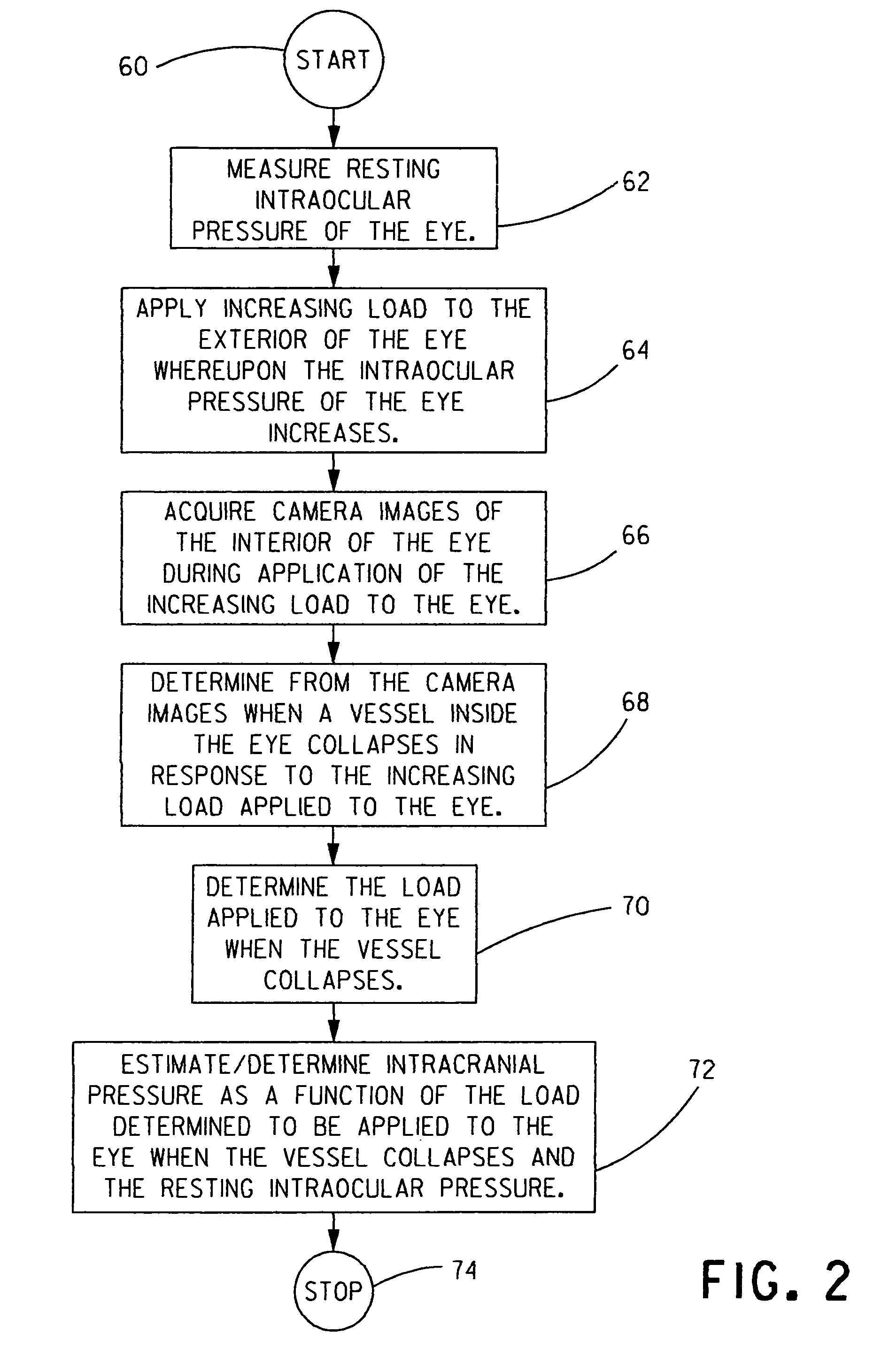
[0034] The controller can automatically determine when the vessel collapses by comparing two or more of the acquired images and determining from said comparison when a reduction in the amount of blood volume in the vessel occurs.

Problems solved by technology

These apparatus and the surgical procedures required for their invasive insertion have many untoward side effects such as bleeding, infection, malfunction, and herniation that may result in permanent disability or death.
Other proposed non-invasive methods and apparatus to measure ICP have not been adapted to medical practice due to practical limitations preventing their use in real world clinical practice.
However, this method is limited both by the fact that a significant percent of the normal population lack this CSF communication due to a normal anatomical variation and by the indirect nature of otoacoustic emission measurement.
This method also requires an intact skull, making it impractical for patients who have skull fractures or surgical opening of the skull during brain surgery.
However, this technique has limited practical use because of the unpredictable nature of the brain's cerebrovascular autoregulatory mechanisms.
The relevance of the measurement disclosed in the Borchert et al. patent is doubtful because onset of papilledema may be delayed 2 to 4 hours after the onset of high ICP.
This deficiency is clinically significant because as ICP increases, cerebral perfusion decreases, leading to decreased brain oxygenation and metabolism.
This 2-4 hour delay can lead to preventable brain injury or even death.
Additionally, a significant percentage of patients with documented ICP elevation lack the ostensible changes in the optic nerve that the technique disclosed in the Borchert et al. patent seeks to identify.
Current methods of ophthalmodynamometry using traditional direct or indirect fundoscopy require a high level of technical training to successfully perform and are subject to inter-observer variability.

Method used


Image

  • Apparatus and method for non-invasive measurement of intracranial pressure
  • Apparatus and method for non-invasive measurement of intracranial pressure
  • Apparatus and method for non-invasive measurement of intracranial pressure

Examples


Embodiment Construction

[0043] The present invention will be described with reference to the accompanying figures.

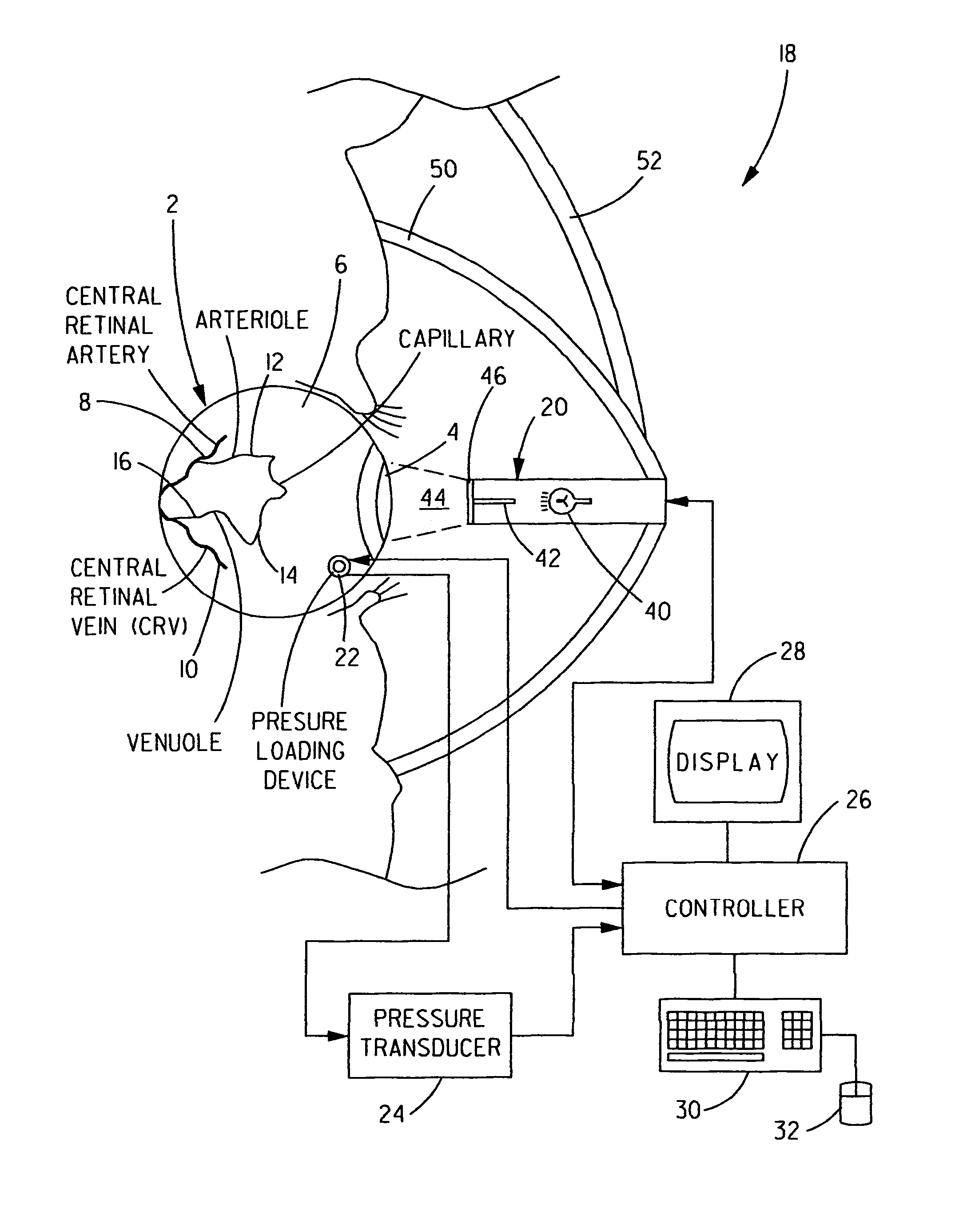
[0044] Within a human eye, the optic nerve travels through the cerebral spinal fluid (CSF) space before entering the interior of the eye. There are two major vessels that run in the optic nerve sheath, namely, a high-pressure central retinal artery and a low-pressure central retinal vein. Other vessels, such as arterioles, capillaries and venuoles, are tributaries of the central retinal artery and the central retinal vein in the eye. The pressure in the central retinal vein (CRV) must be greater than the intracranial pressure (ICP) surrounding the optic nerve sheath in order for blood to flow through the optic nerve sheath. The pressure required to collapse the CRV, called the venous outflow pressure (VOP), may be used to determine ICP.

[0045] With reference to FIG. 1, a human eye 2 includes a cornea 4 and a sclera 6. An interior of eye 2 includes a central retinal artery 8, a central retinal ...


PUM


Abstract

An intracranial pressure (ICP) within a patient's skull can be determined by observing a vessel in the patient's eye, desirably in the red and / or infrared (IR) spectrum, while causing the pressure inside the eye to increase. On or about the time the observed vessel collapses in response to increasing the pressure inside the eye, the pressure inside the eye is determined. The ICP can then be determined as a function of the pressure inside the eye. Desirably, the vessel being observed is the central retinal vein of the eye.

Description

CROSS REFERENCE TO RELATED APPLICATIONS [0001] The present application claims priority from U.S. Provisional Application No. 60 / 656,449, filed Feb. 24, 2005, and U.S. Provisional Patent Application No. 60 / 703,391, filed Jul. 29, 2005, both of which are incorporated herein by reference.BACKGROUND OF THE INVENTION [0002] 1. Field of the Invention [0003] The present invention relates to determining intracranial pressure in patients and, more particularly, to determining intracranial pressure by non-invasively determining the point when one or more vessels with the eye of a patient collapses when a known load is applied to the exterior of the eye. [0004] 2. Description of Related Art [0005] Intracranial pressure (ICP) is an important parameter in the management of conditions such as traumatic brain injury, stroke, intracranial hemorrhage, central nervous system (CNS) neoplasm, CNS infections and hydrocephalus where cerebral edema exists or brain compliance is altered. High ICP must be a...

Claims


Application Information

Patent Timeline
IPC IPC(8): A61B5/00A61B5/04
CPCA61B3/12A61B5/031
Inventor BRAXTON, ERNEST E.
Owner BRAXTON ERNEST E
Learn More