Surgical tool and method for extracting tissue from wall of an organ

a tissue extraction and surgical technology, applied in the field of surgical tools and methods, can solve the problems of buttonholing or penetrating vital tissues, affecting the function of the organ, and not being able to control the symptoms of glaucoma with medication,

Inactive Publication Date: 2006-10-12
D O T & OPHTHALMIC TECH
View PDF2 Cites 11 Cited by
  • Summary
  • Abstract
  • Description
  • Claims
  • Application Information

AI Technical Summary

Benefits of technology

[0036] According to a further feature of the present invention, the second blade element is restrained from pivotal movement when the second blade assembly assumes the closed position.
[0039] According to a further feature of the present invention, the first blade assembly and the second blade assembly are configured such that, when in the closed position, the first blade element and the second blade element are restrained against pivotal movement.

Problems solved by technology

However, medications often fail to control some forms of glaucoma.
This surgery encounters many complications, ranging from loss of the volume and shape of the eye when the fistula is too large through to formation of scar tissue which seals down the fistula.
The latter requires peri-operative application or injection of anti-metabolites, harboring a significant risk of bleeding, infections, wound leaks, buttonholing or penetrating vital tissues.
While this approach should theoretically offer advantages of minimizing complications due to damage to the conjunctiva, in practice, the results have been poor, primarily due to complications associated with the typically large corneal incision.

Method used


Image

  • Surgical tool and method for extracting tissue from wall of an organ
  • Surgical tool and method for extracting tissue from wall of an organ
  • Surgical tool and method for extracting tissue from wall of an organ

Examples


Embodiment Construction

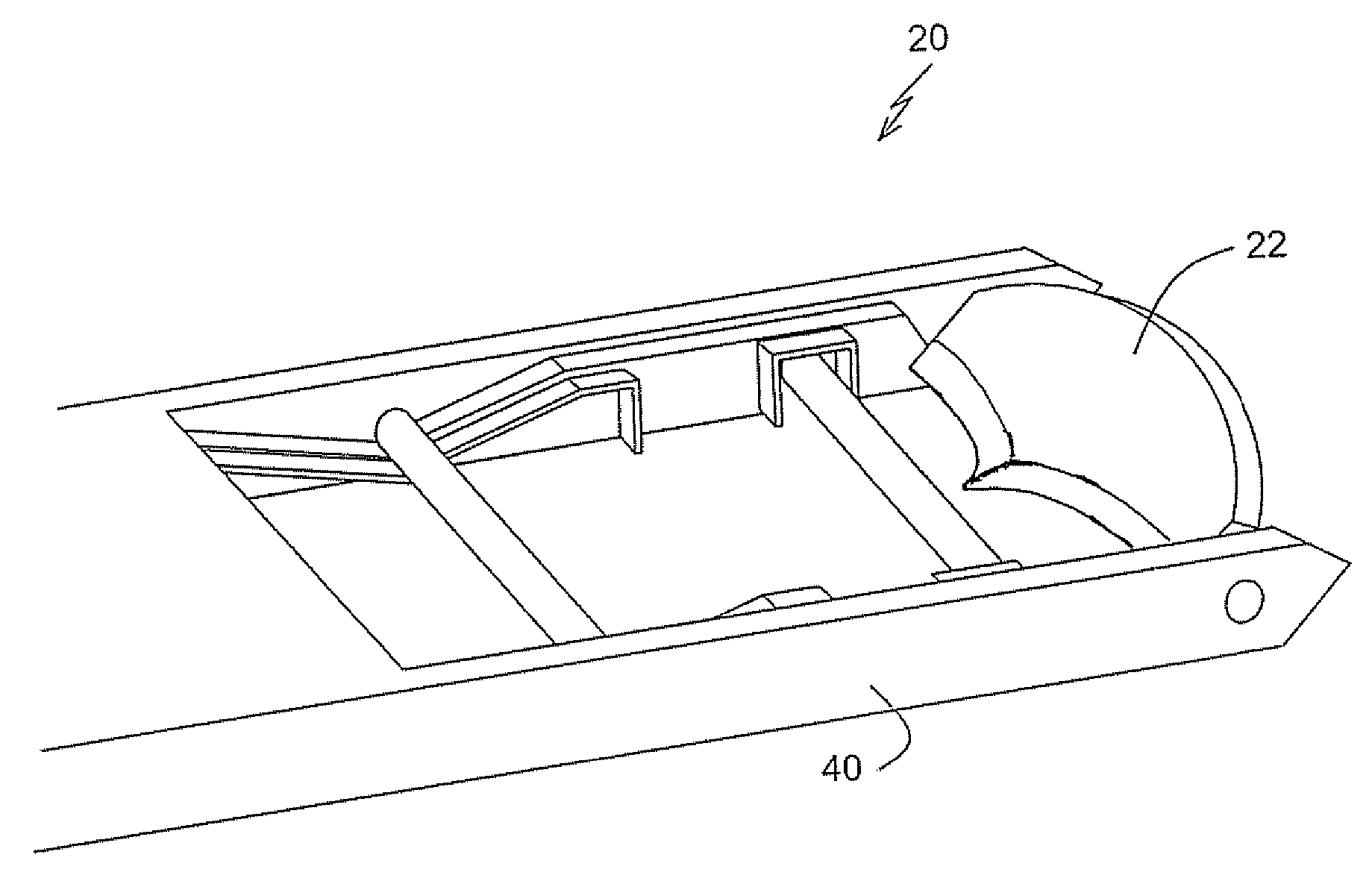
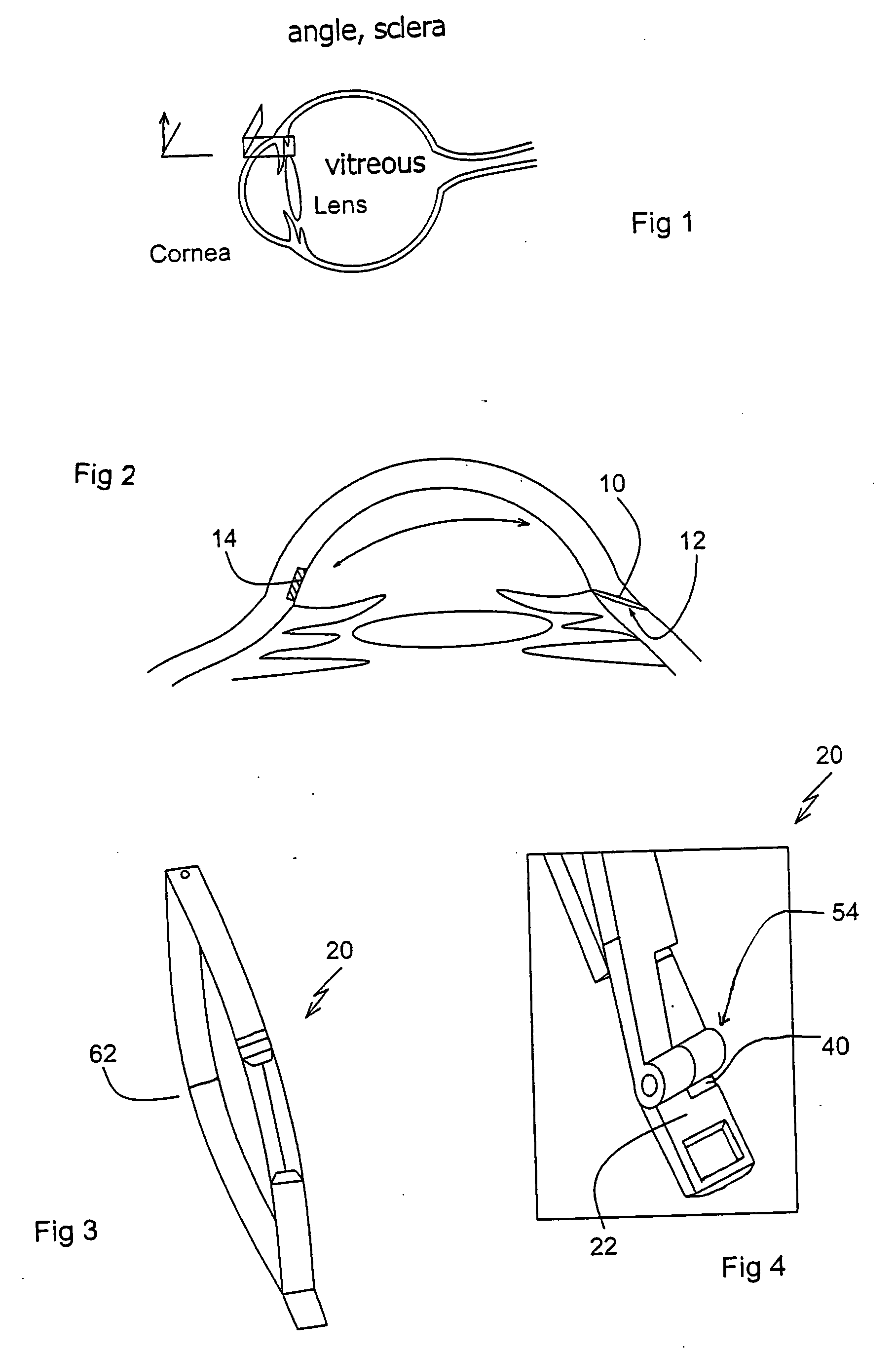
[0070] The present invention is a method for extracting a tissue block from the wall of a hollow organ and forming a self-sealing flap in tissue of the wall. Also provided is a preferred example of a surgical tool and a corresponding specific example of implementation of the method of the invention.

[0071] The principles and operation of surgical tools and methods according to the present invention may be better understood with reference to the drawings and the accompanying description.

[0072] Referring now to the drawings, FIG. 2, an enlarged view of a part of the eye shown in FIG. 1, shows the underlying principles of a surgical method for extracting a tissue block from the wall of a hollow organ and forming a self-sealing flap in tissue of the wall, particularly as applied to the eye. Thus, in general terms, the method includes forming an elongated slit 10 of substantially constant width extending from an outer surface of the wall into the wall, typically at a shallow angle, so a...


PUM


Abstract

The present invention is concerned with methods and equipment for the treatment of the eye and more particularly with an instrument to increase eye filtration and treat glaucoma. The instrument is surgical tool operated entirely by the surgeon's hands and is composed of a hand-piece and an eye-penetrating intraocular segment each having two opponent and reciprocally operating fork-like arms and blade-sharp leaves, respectively. The eye-penetrating segment is a rigid embodiment of a size to create a limbus-pointing tunnel in the cornea, penetrate the eye of a patient through a water tight incision and, in a five step dissect and extract procedure, remove from within the eye a trabecular tissue block leaving the incision to self-seal by intraocular pressure. In the same “modus operandi”, a sample tissue can be extracted from any other hollowed or parenchimatous organ.

Description

[0001] This application is a Divisional of U.S. Ser. No. 10 / 135,365 filed May 1, 2002, currently pending.FIELD AND BACKGROUND OF THE INVENTION [0002] The present invention relates to surgical tools and methods and, in particular, it concerns a surgical tool and corresponding method for extracting tissue from wall of an organ. The tool and method are particularly suited to procedures for lowering intraocular pressure. [0003] Glaucoma is a disease in which intraocular pressure is a causative risk factor. It affects significant numbers of our population. The treatment of glaucoma is usually medicinal to lower the intraocular pressure. However, medications often fail to control some forms of glaucoma. When further treatment is required, a microsurgical operative procedure is performed. [0004] Conventional surgical techniques for intraocular pressure reduction approach the tissue in the angle of the eye through the conjunctiva. The procedure involves constructing a fistula or opening in ...

Claims


Application Information

Patent Timeline
Patent Type & Authority Applications(United States)
IPC IPC(8): A61F9/00A61F9/007
CPCA61F9/0133A61F9/00781
Inventor DAN, JACOB
Owner D O T & OPHTHALMIC TECH
Why Eureka
Try Eureka