
System and method for using learned discriminative models to segment three dimensional colon image data

a three-dimensional colon image and discriminative model technology, applied in image enhancement, image analysis, instruments, etc., can solve the problems of difficult to distinguish colon walls from colon walls, small polyps of interest, and difficult task of automatic segmentation

Active Publication Date: 2006-10-26
SIEMENS HEALTHCARE GMBH
View PDF7 Cites 18 Cited by
  • Summary
  • Abstract
  • Description
  • Claims
  • Application Information

AI Technical Summary

Benefits of technology

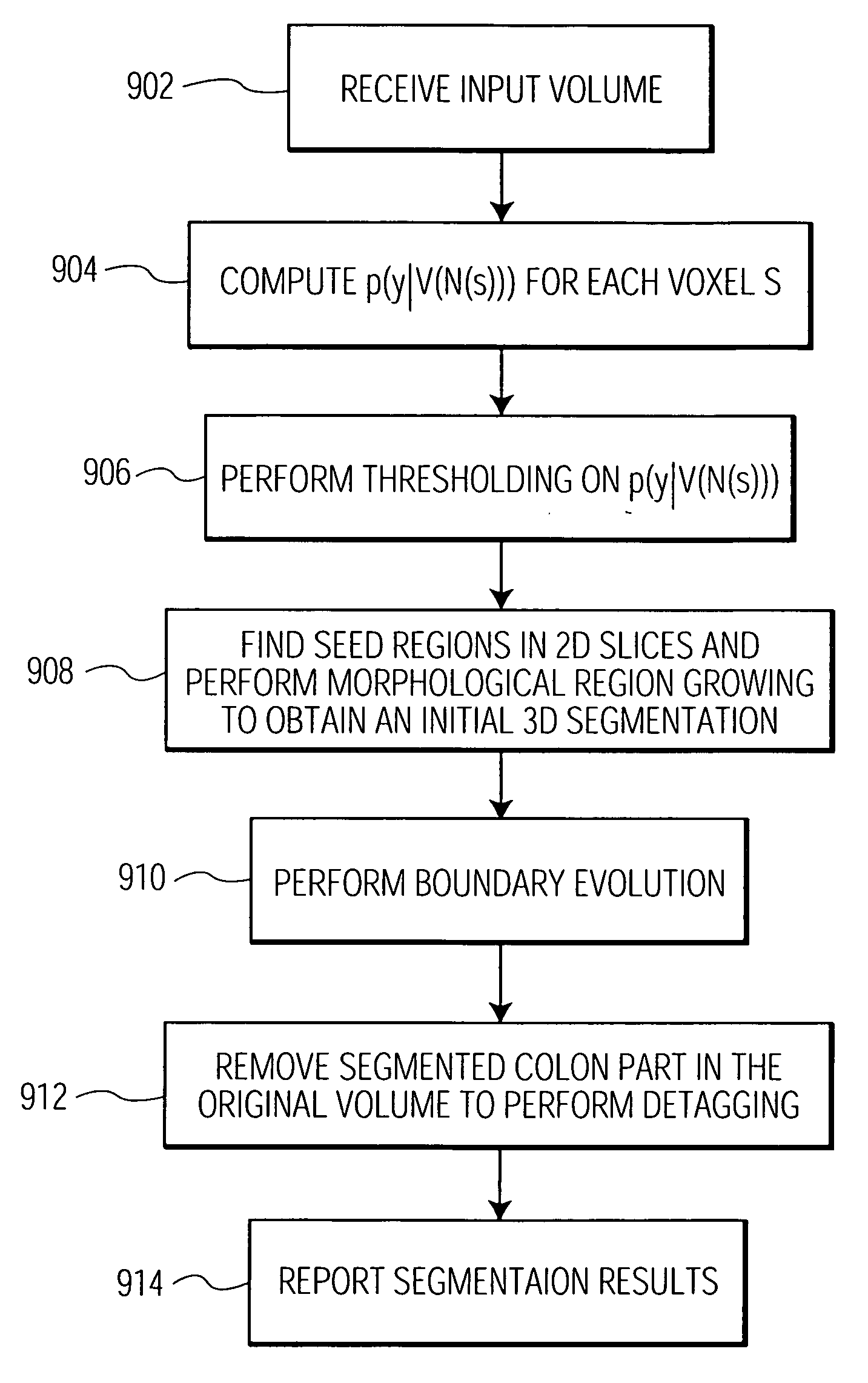
The present invention is a system and method for segmenting a border of an anatomical structure in a 3D image using learned discriminative models. The system calculates a discriminative probability model for each voxel in the image and performs thresholding on these models. The system then obtains 2D slices along X-Y planes and selects seed regions in these slices. The system performs morphological region growing on these selected seed regions to obtain an initial 3D segmentation. The system then performs boundary evolution on this initial segmentation to remove the segmented anatomical structure from the original 3D image. The technical effect of this invention is to provide a reliable and accurate method for segmenting the border of an anatomical structure in a 3D image.

Problems solved by technology

This is very inconvenient and prevents virtual colonoscopy from being a general screening tool for a large population.
The task of automatic segmentation is very challenging.
Second, polyps of interest are very small and don't have unique intensity patterns, nor have any special shapes.
It is hard to distinguish them from the colon wall, especially when they are surrounded by tagged material.
Third, the volumetric data to be processed is massive (e.g., 400×512×512), which eliminates the possibility of using any computationally expensive method.
This becomes essentially a segmentation problem in which the task is to delineate and locate the colon wall.
However, residual materials observe large variation in appearance depending upon where they are, what the patient eats, and how much they are tagged.
Furthermore, the challenge of segmenting an uncleansed colon comes from the fact that residual materials and colon walls observe complex patterns, which are hard to separate.
This is representative of a large class of problems in medical imaging and vision in which the task is to perform foreground / background segmentation.
However, these models, such as Gaussian, only work in very constrained situations since it is difficult to use them to capture the larger intra-class variability and inter-class similarity.

Method used


Image

  • System and method for using learned discriminative models to segment three dimensional colon image data
  • System and method for using learned discriminative models to segment three dimensional colon image data
  • System and method for using learned discriminative models to segment three dimensional colon image data

Examples


Embodiment Construction

[0020] The present invention is directed to a system and method for using learned discriminative models to detect the appearance of complex foreground and background in an image using a probabilistic boosting tree and boundary evolution. Such a method is particularly effective in the segmentation and delineation of a colon border in 3D colon image data. In accordance with one embodiment of the present invention, residual material (e.g., stool) is segmented from the colon wall by tagging the residual material. The tagged residual material is given a high intensity so that it shows up as bright areas in the image. A learning based method is then used to determine the presence of the colon border.

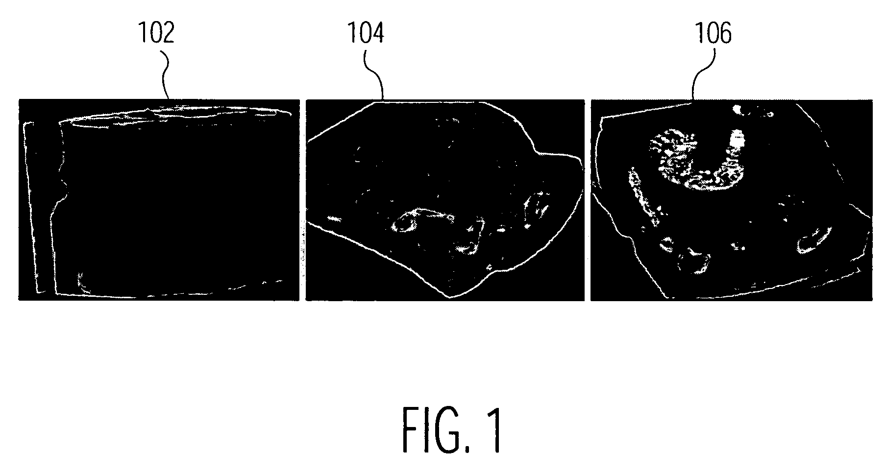
[0021] Such image data can be obtained using different imaging modalities such as Computed Tomography (CT), X-ray or Magnetic Resonance Imaging (MRI). FIG. 1 illustrates a number of views of a colon. The first image 102 shows a complete CT volume of a section of a colon. There are two types o...


PUM


Abstract

A system and method for using learned discriminative models to segment a border of an anatomical structure in a three dimensional (3D) image is disclosed. A discriminative probability model is computed for each voxel in the 3D image. Thresholding is performed on each discriminative probability model. One or more two dimensional (2D) slices of the thresholded 3D image along X-Y planes are obtained. Seed regions are selected in the 2D slices. Morphological region growing is performed on the selected seed regions. An initial 3D segmentation is obtained. Boundary evolution is performed on the initial 3D segmentation. The segmented anatomical structure is removed. in the original 3D image.

Description

CROSS REFERENCE TO RELATED APPLICATIONS [0001] This application claims the benefit of U.S. Provisional Application Ser. No. 60 / 652,280 filed on Feb. 10, 2005, and U.S. Provisional Application Ser. No. 60 / 660,136, filed on Mar. 9, 2005 which are incorporated by reference in their entirety.FIELD OF THE INVENTION [0002] The present invention is directed to a system and method for segmenting an anatomical structure in a three dimensional image, and more specifically, to a system and method for segmenting three dimensional colon image data by using learned discriminative models to delineate foreground and background image data. BACKGROUND OF THE INVENTION [0003] Colon cancer is one of the leading causes of death in the U.S. The number of deaths can be largely reduced if polyps can be detected and treated at their early stage of development. Virtual colonoscopy is a new technology being developed to help doctors find polyps in three dimensional (3D) computed tomography (CT) image data. Ho...

Claims


Application Information

Patent Timeline
Patent Type & Authority Applications(United States)
IPC IPC(8): G06K9/34G06V10/28
CPCG06K9/38G06K9/4638G06K2209/05G06T7/0081G06T2207/30028G06T2207/10081G06T2207/20081G06T2207/20156G06T2200/04G06T7/11G06V10/28G06V10/457G06V2201/03
Inventor TU, ZHUOWENZHOU, XIANGCOMANICIU, DORINBOGONI, LUCABARBU, ADRIAN
Owner SIEMENS HEALTHCARE GMBH
Learn More