Eureka AIR delivers breakthrough ideas for toughest innovation challenges, trusted by R&D personnel around the world.

System for processing medical image representative data from multiple clinical imaging devices

Inactive Publication Date: 2006-10-26
SIEMENS MEDICAL SOLUTIONS USA INC
View PDF15 Cites 77 Cited by
  • Summary
  • Abstract
  • Description
  • Claims
  • Application Information

AI Technical Summary

Problems solved by technology

In existing systems report generation typically begins after image data acquisition, which is time consuming with multiple actors reproducing the same information.
This may result in a clinician failing to see multiple small “anomalies” occurring in images derived from multiple corresponding different imaging modalities (such as MR, CT, X-ray, Ultrasound etc.) that individually may be missed by a clinician.
Existing system also are inefficient in enabling a user to locate and display selected data.

Method used


Image

  • System for processing medical image representative data from multiple clinical imaging devices
  • System for processing medical image representative data from multiple clinical imaging devices
  • System for processing medical image representative data from multiple clinical imaging devices

Examples


Embodiment Construction

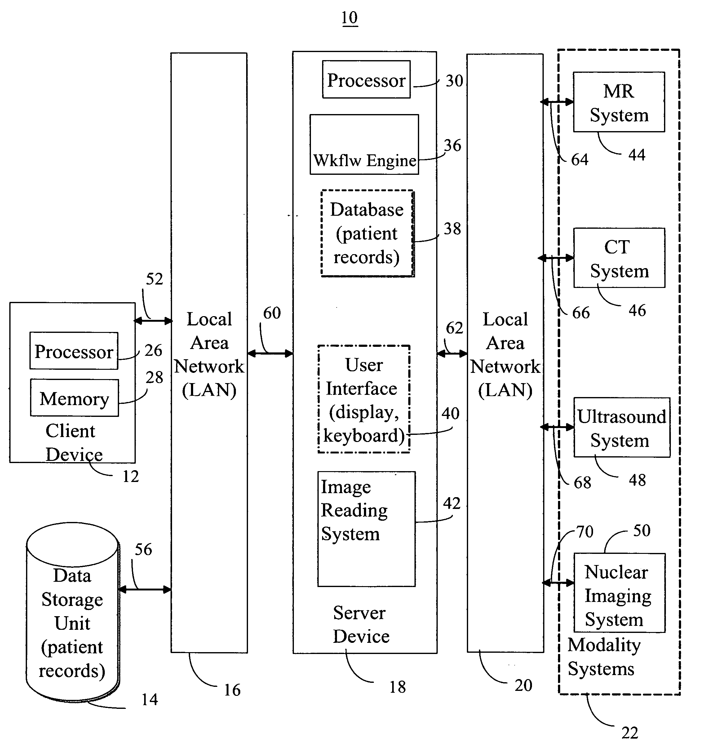
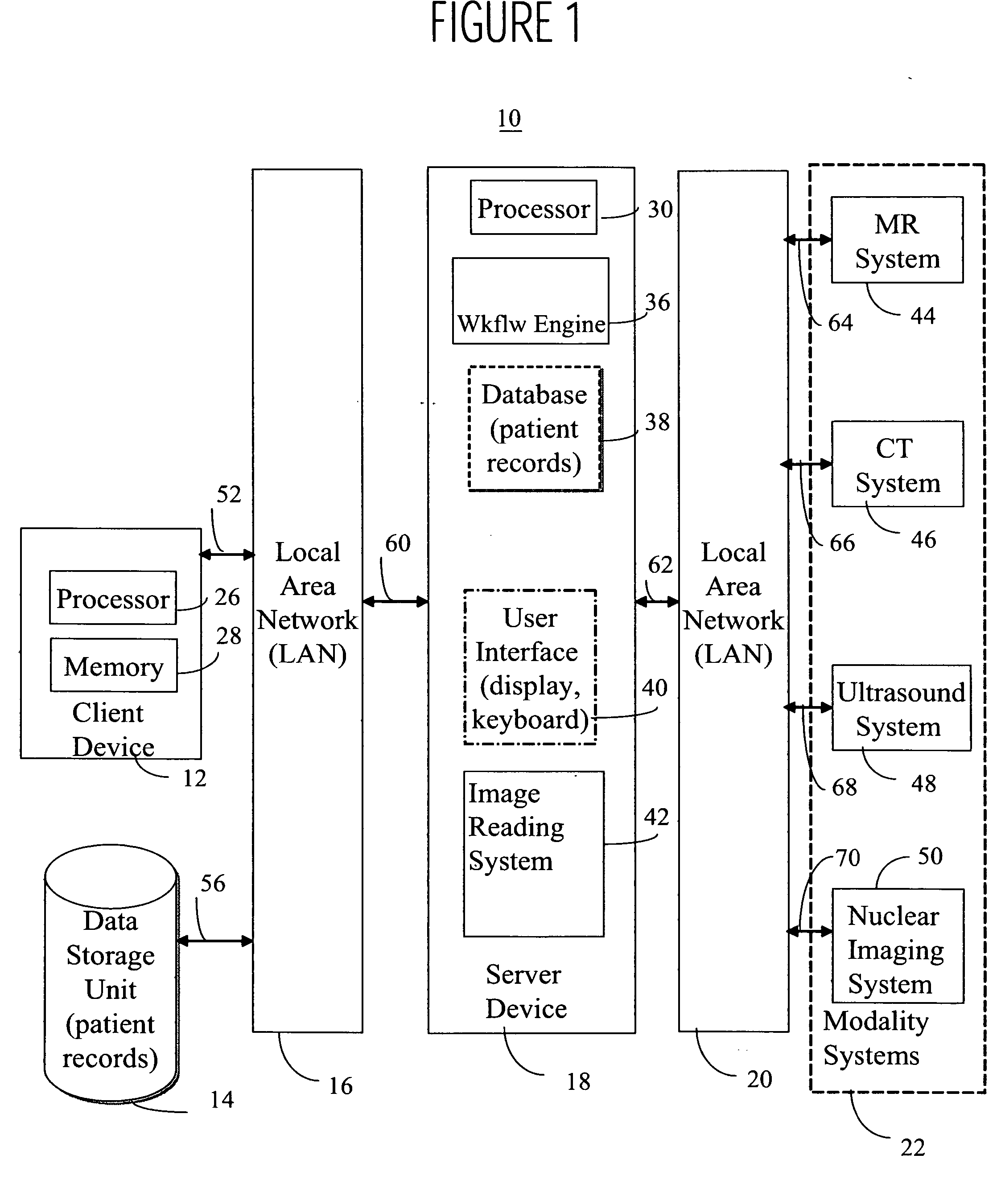
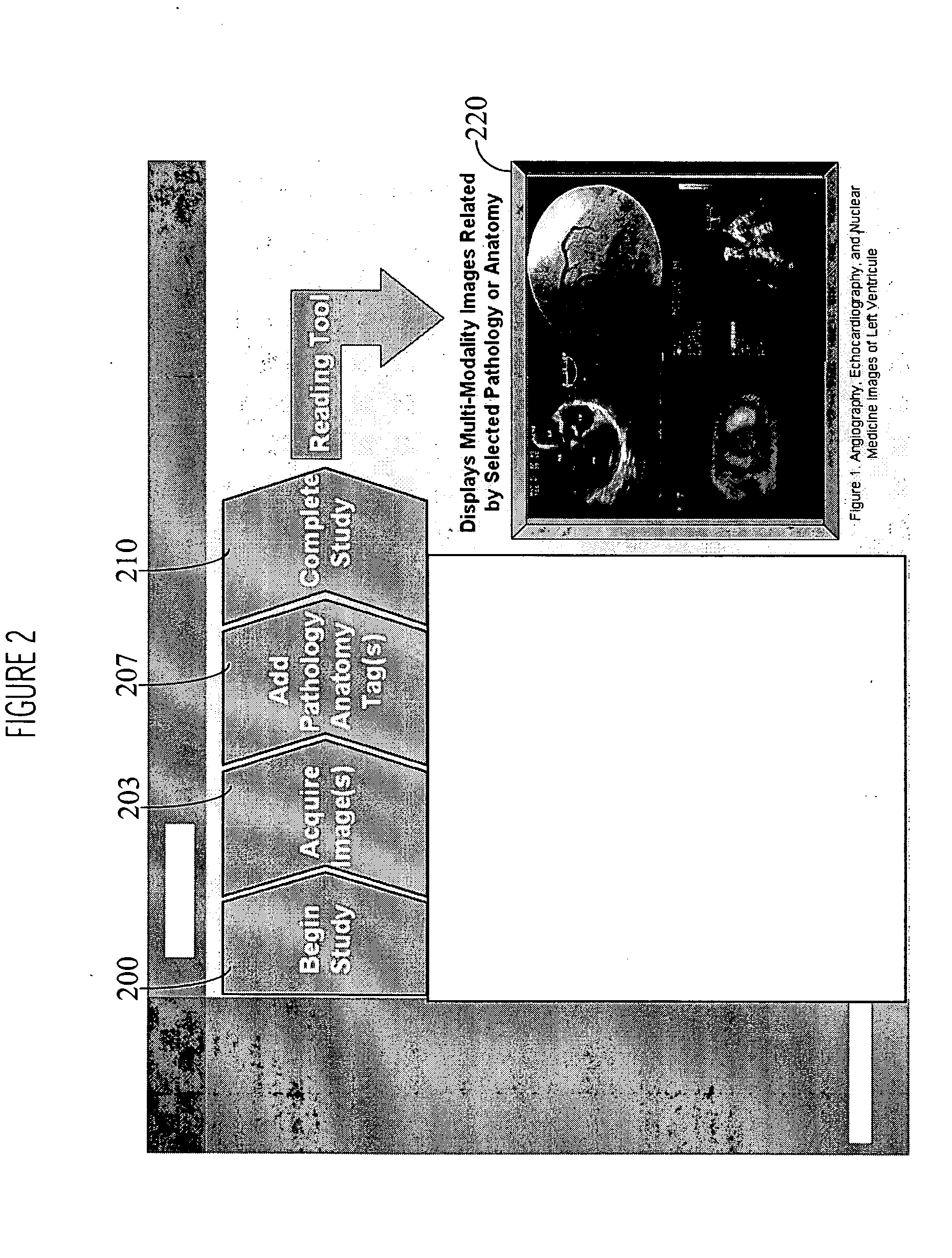
[0010]FIG. 1 shows a hospital information network 10 including a multi modality image reading system 42. The system incorporates a workflow engine 36 to support report generation earlier in a workflow cycle than in a typical existing image reading system. Image reading system 42 associates related images of a particular patient derived from multiple different modality imaging devices such as MR, CT, X-ray, Ultrasound, etc. Multi-modality image reading system 42 associates images derived from multiple different modality devices based on pathology and on anatomic layout in order to advantageously provide a user with an overall comprehensive clinical view of relevant patient medical image data. In a preferred embodiment image reading system 42 also generates a template (framework) of a report during image data acquisition rather than following image data acquisition. Image reading system 42 employs user interface system 40 including a configuration processor enabling a user to assign t...


PUM


Abstract

A multi imaging modality reading system enables medical report generation to begin earlier in a workflow process by enabling automatic image correlation of images derived by one or multiple, imaging modalities allowing for comparison of pathology over time and between modalities. A user interface system for accessing multiple medical images derived from different types of medical imaging systems includes at least one repository. The at least one repository associates, multiple different medical images derived from corresponding multiple different types of medical imaging systems, with data identifying a particular anatomical body part of a particular patient and with data identifying the different types of medical imaging systems. A display processor accesses the at least one repository and initiates generation of data representing a composite display image including multiple image windows individually including different medical images derived from corresponding multiple different types of medical imaging systems for a particular anatomical body part of a particular patient.

Description

[0001] This is a non-provisional application of provisional application Ser. No. 60 / 653,789 by M. Esham filed Feb. 17, 2005.FIELD OF THE INVENTION [0002] This invention concerns a user interface system for accessing multiple medical images derived from different types of medical imaging systems. BACKGROUND OF THE INVENTION [0003] In existing medical imaging report generation systems, a user is typically required to formulate time-consuming reports following image data acquisition and to view multiple exams as separate imaging studies, therefore requiring the user to manually compile and integrate the information into a single knowledge-view. In existing systems report generation typically begins after image data acquisition, which is time consuming with multiple actors reproducing the same information. This may result in a clinician failing to see multiple small “anomalies” occurring in images derived from multiple corresponding different imaging modalities (such as MR, CT, X-ray, U...

Claims


Application Information

Patent Timeline
IPC IPC(8): G06F17/30G16H30/20G16H30/40
CPCG06F19/3487G06F19/321G16H15/00G16H30/40G16H30/20
Inventor ESHAM, MATTHEW PAULRICHTER, MELISSAPOYNTON, RICHARD
Owner SIEMENS MEDICAL SOLUTIONS USA INC
Why Eureka
Eureka Blog
Learn More