A Quantification and Extraction Method of Vessel Wall Image Texture Features

An image texture and extraction method technology, applied in catheters and other directions, can solve the problems of inability to distinguish vascular lesions, application, popularization influence, and high operator requirements, and achieve the effect of improving functions, facilitating clinical promotion, and easy acceptance.

Active Publication Date: 2016-02-24
NAT INST OF ADVANCED MEDICAL DEVICES SHENZHEN
View PDF6 Cites 0 Cited by
  • Summary
  • Abstract
  • Description
  • Claims
  • Application Information

AI Technical Summary

Problems solved by technology

However, because of its cumbersome operation and high requirements for operators, its clinical application and popularization are affected.
Therefore, these detection methods are usually unable to effectively distinguish vascular lesions in the early stage when the vascular structure and function have not changed.

Method used


Image

  • A Quantification and Extraction Method of Vessel Wall Image Texture Features
  • A Quantification and Extraction Method of Vessel Wall Image Texture Features
  • A Quantification and Extraction Method of Vessel Wall Image Texture Features

Examples


Embodiment Construction

[0026] The present invention will be described in further detail below in conjunction with the accompanying drawings and specific embodiments.

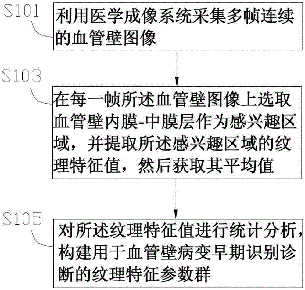
[0027] see figure 1 , an embodiment of the present invention provides a method for quantifying and extracting texture features of a blood vessel wall image, which includes the following steps:

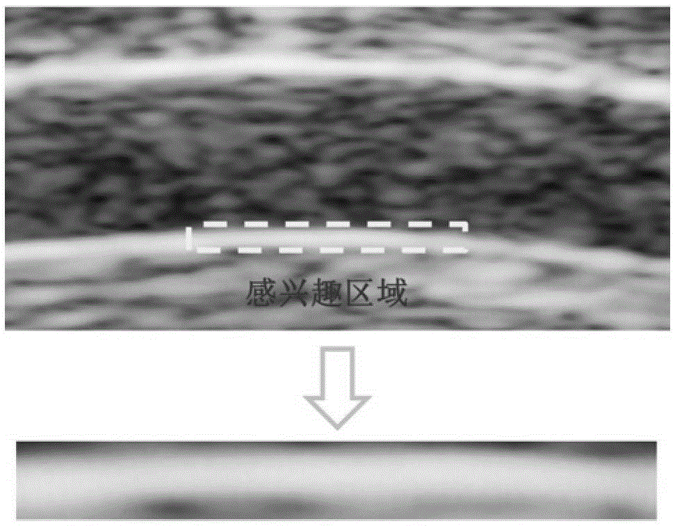
[0028] Step S101, using a medical imaging system to acquire multiple frames of continuous blood vessel wall images.

[0029] In this embodiment, a medical imaging system is used to collect N frames of continuous blood vessel wall images, and N is an image covering at least one cardiac cycle. It is 60Hz / min, cardiac cycle Tc=60 / f=1 second; then N=m×FR×Tc=100m frames (m=1, 2, 3...). That is, N should be an integer multiple of 100, such as figure 2 shown.

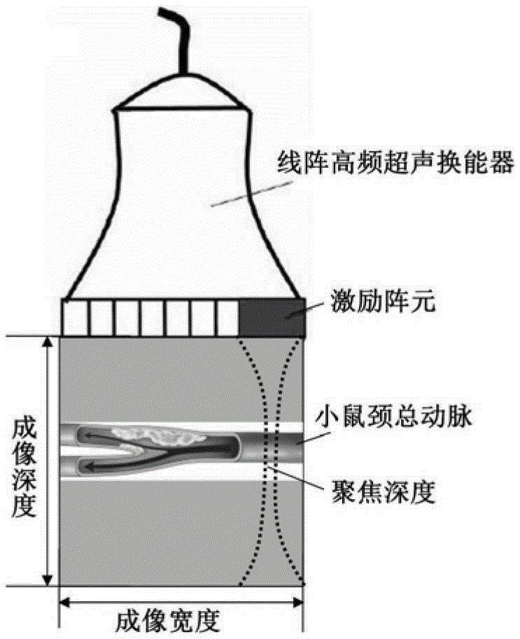
[0030] It can be understood that the medical imaging system may be an ultrasound imaging system, an optical imaging system (such as an X-ray machine), a CT imaging system, or an MRI imagi...


PUM


Abstract

The invention provides a method for quantifying and extracting image texture characteristics of vascular wall and is used for early recognition and diagnosis of vascular wall pathological changes. The method includes steps of acquiring vascular wall images of multiple continuous frames by means of a medical imaging system; selecting an inner membrane-intermediate membrane layer of the vascular wall as an interested area from the vascular wall images of each frame, extracting texture characteristic values of the interested area, and acquiring average value of the texture characteristics values; and computing and analyzing the texture characteristic values and establishing a texture characteristic parameter group used for early recognition and diagnosis of the vascular wall pathological changes. The method for quantifying and extracting image texture characteristics of the vascular wall is capable of extracting the texture characteristic values of the vascular wall in any interested area, is beneficial to accurate positioning of pathological changes during pathological changes detection of the vascular wall, high in applicability, free of hardware upgrading in existing clinical imaging system, low in cost, easy to accept by hospitals and doctors and convenient to clinically popularize.

Description

technical field [0001] The invention relates to a method for quantifying and extracting texture features of blood vessel wall images, in particular to a method for quantifying and extracting texture features of blood vessel wall images based on image texture features. Background technique [0002] Cardiovascular and cerebrovascular diseases seriously endanger human health and have become an increasingly urgent major social public problem. Atherosclerosis is the chief culprit of cardiovascular and cerebrovascular diseases. Atherosclerosis is a long-term and complicated process. If it can be accurately identified and diagnosed in the early stage, timely intervention and treatment can be carried out in the early stage of the disease, which can effectively improve the cure rate and reduce medical expenses. At present, the main clinical basis for judging atherosclerosis is vascular structure, function and hemodynamic factors, that is, intima-media thickness, presence or absence ...

Claims


Application Information

Patent Timeline
Patent Type & Authority Patents(China)
IPC IPC(8): A61B5/02
Inventor 郑海荣牛丽丽钱明孙风雷肖扬王丛知
Owner NAT INST OF ADVANCED MEDICAL DEVICES SHENZHEN
Why Eureka
Try Eureka