
Method and apparatus for human brain imaging using a nuclear medicine camera

a nuclear medicine camera and brain imaging technology, applied in the field of nuclear medical imaging arts, can solve the problems of patient's shoulders affecting the detector, the radioactivity of radiopharmaceuticals is limited by permissible levels, and the inability to detect the brain, etc., to achieve the effect of improving signal sensitivity and image resolution

Inactive Publication Date: 2006-11-23
KONINKLIJKE PHILIPS ELECTRONICS NV
View PDF10 Cites 19 Cited by
  • Summary
  • Abstract
  • Description
  • Claims
  • Application Information

AI Technical Summary

Benefits of technology

[0017] Another advantage resides in facilitating studies of constricted anatomical regions such as the head, neck, or limbs using gamma cameras with large-area detector heads.

Problems solved by technology

For nuclear medical imaging, the radioactivity of the radiopharmaceutical is limited by permissible levels of patient radiation exposure.
The combination of a close conformal path and relatively large-area detector heads can make tomographic SPECT imaging of constricted anatomical regions such as the head, neck, or limbs problematic.
In the case of tomographic head or neck imaging, for example, the patient's shoulders can interfere with the detector head or with a mounting stricture supporting the detector head.
However, this approach has been found to compromise image resolution.
These techniques are not wholly satisfactory for tomographic imaging of constricted anatomical regions.
In other trajectory portions, the choice of zoom may not be beneficial, and indeed may even be detrimental.
This imposes further compromises upon selection of the zoom area, since an optimal zoom area for one detector may be non-optimal for the opposing detector.

Method used


Image

  • Method and apparatus for human brain imaging using a nuclear medicine camera
  • Method and apparatus for human brain imaging using a nuclear medicine camera
  • Method and apparatus for human brain imaging using a nuclear medicine camera

Examples


Embodiment Construction

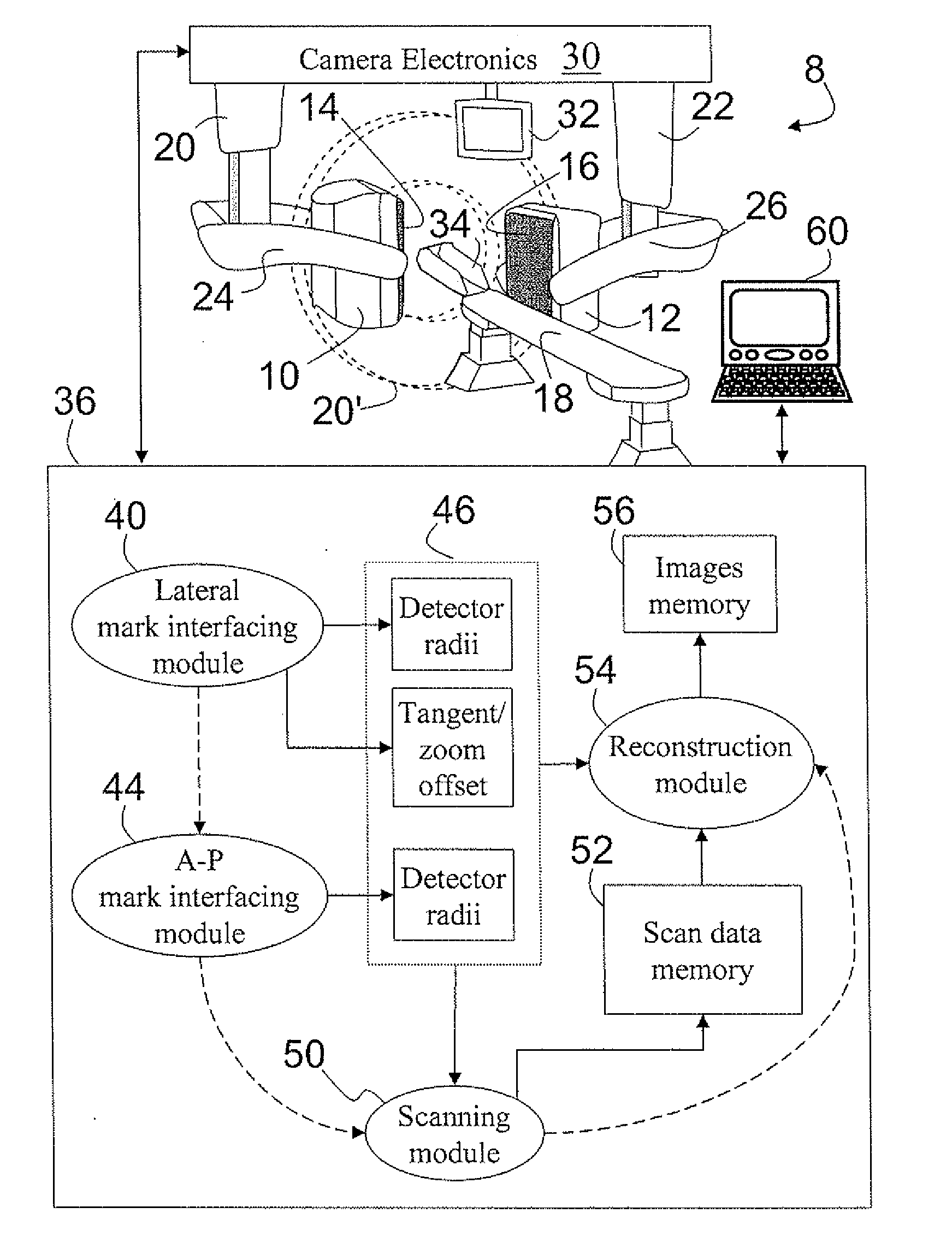
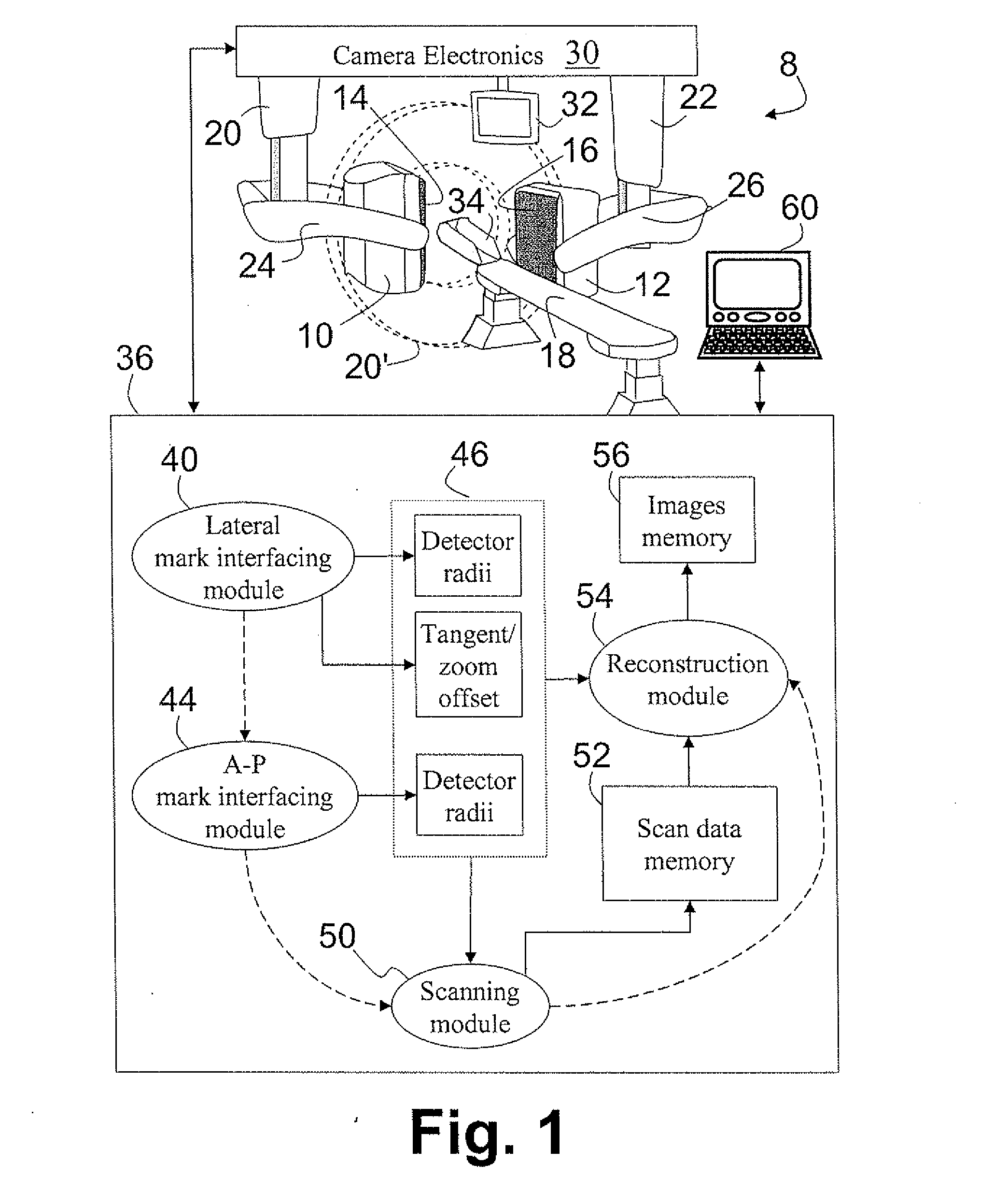
[0026] With reference to FIG. 1, a nuclear medical imaging system includes a gamma camera 8, which is a two-detector head camera having first radiation detector head 10 and second radiation detector head 12. The radiation detector heads 10, 12 have radiation-sensitive faces 14, 16, respectively, which in FIG. 1 are generally arranged to face a patient support or couch 18. Although not shown at the level of detail of FIG. 1, in typical embodiments the radiation-sensitive faces 14, 16 each include a honeycomb collimator (optionally detachable and replaceable to enable a selection of collimation characteristics), a scintillator arranged to receive the collimated radiation, and photomultiplier tubes (PMTs), photodiodes, or other optical detectors arranged to detect the scintillations. However, the radiation-sensitive faces 14, 16 can employ other radiation detection technologies, such as solid-state CZT-based detectors. Moreover, the number of detector heads can be one or can be greater...


PUM


Abstract

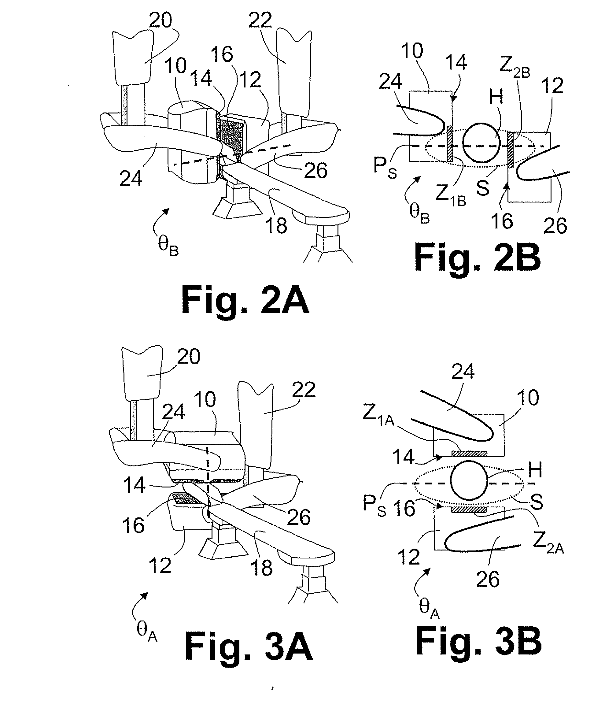
In an imaging method, mark positions are defined for one or more detector heads (10, 12) at one or more marked angular orientations (θA, θB). The mark positions for at least one marked angular orientation (θB) include a tangential offset of at least one detector head. Imaging data are acquired using the one or more detector heads following a conformal trajectory passing through the defined mark positions. The acquired imaging data are reconstructed into a reconstructed image.

Description

BACKGROUND [0001] The following relates to the nuclear medical imaging arts. It particularly relates to head, neck, and brain scans performed using single-photon emission computed tomography (SPECT) cameras, and will be described with particular reference thereto. However, the following relates more generally to imaging of constricted anatomical regions such as the head, neck, or limbs using movable detector heads that follow trajectories closely conforming with the outer dimensions of the imaged anatomy. [0002] In nuclear medical imaging techniques such as SPECT, a radiopharmaceutical is administered to the patient or other imaging subject. The radiopharmaceutical is typically designed to preferentially collect in an organ or tissue type of interest. For example, an intravenously administered radiopharmaceutical that remains in the blood system can be used to image patient vasculature, or the radiopharmaceutical can be designed to collect in preselected brain tissue to measure its ...

Claims


Application Information

Patent Timeline
IPC IPC(8): G01T1/166
CPCG01T1/1648A61B6/4458A61B6/4452
Inventor D'AMBROSIO, RAYMOND C.GARRARD, JODY L.KARMALAWY, MOATAZ
Owner KONINKLIJKE PHILIPS ELECTRONICS NV
Learn More